Numbers of MRI

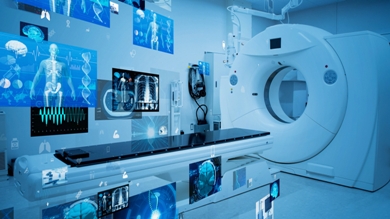
During an MRI scan, patients are typically placed inside a cylindrical machine and exposed to a combination of radio frequency pulses and electromagnetic fields. These signals create an overall picture of the body’s internal structures, which can be interpreted by specialized software or medical professionals. Though the scan itself is relatively simple, there are certain steps that must be taken before and after the procedure in order to ensure patient safety. For example, subjects must remove all metallic objects such as watches or jewelry before entering the MRI machine..


Radiology plays a crucial role in diagnosing and treating various medical conditions. Whether you need an X-ray, ultrasound, CT scan, or MRI, finding the best radiologist near you is essential for accurate results and effective treatment. If you are searching for "best radiologist in Pune" or "radiology centre in Pune," it’s important to choose a center that offers high-quality services, experienced radiologists, and state-of-the-art technology.
A radiologist is a medical professional who uses imaging techniques to diagnose and treat diseases. From routine check-ups to critical diagnostics, the right radiologist can make all the difference in ensuring that you receive the best care possible. When looking for "radiology near me" or "radiology centre near me," it’s vital to choose a clinic that has a reputation for accuracy, precision, and patient care. The best radiologists in Pune are skilled in interpreting complex imaging results and providing the necessary guidance for your treatment.
If you are looking for affordable and high-quality radiology services, Diagnopein is your ideal destination. With a team of expert radiologists and advanced diagnostic equipment, Diagnopein offers comprehensive radiology services at competitive prices. Our goal is to make advanced diagnostic services accessible to everyone, ensuring that you get the best results without breaking the bank.
At Diagnopein, we offer a wide range of radiology services, including:
1] X-rays: For diagnosing fractures, infections, and other skeletal issues.
2] Ultrasounds: For evaluating soft tissues, organs, and monitoring pregnancy.
3] CT Scans (CT Imaging): For detailed images of the body’s internal structures, helping diagnose tumors, injuries, and internal conditions.
4] MRI Scans: Providing high-resolution images for accurate diagnosis of neurological, muscular, and skeletal conditions.
If you’re searching for "low-cost radiology services near me," Diagnopein offers affordable diagnostic solutions without compromising quality. We understand that healthcare costs can be a burden, which is why we provide transparent pricing and various payment options to ensure that everyone can access the necessary diagnostic services.
With Diagnopein, you don’t have to worry about hidden charges or unnecessary fees. Our pricing structure is designed to be budget-friendly while still providing high-quality, accurate radiology services.
The radiologists at Diagnopein are highly qualified and experienced in their field. They are dedicated to providing precise interpretations of your scans, ensuring that you get the correct diagnosis. Whether you need a simple X-ray or a complex MRI, our radiologists use the latest technology and their expertise to ensure the most accurate results.
If you're searching for "radiology centre near me" or "radiology centre in Pune," Diagnopein's central location in Pune makes it easy for you to access top-tier diagnostic services. We prioritize convenience, offering flexible hours and a comfortable environment so that you can undergo your tests with minimal stress.
Don’t wait for symptoms to worsen—early detection is key to effective treatment. Whether you need routine imaging or more advanced diagnostics, Diagnopein provides a full range of radiology services at affordable prices. Book your appointment with Diagnopein today and experience the best radiology services in Pune.
Choose Diagnopein for accurate, affordable, and compassionate care that ensures your health is in good hands.
Both MRIs and CT scans can view internal body structures. However, a CT scan is faster and can provide pictures of tissues, organs, and skeletal structure. An MRI is highly adept at capturing images that help doctors determine if there are abnormal tissues within the body. MRIs are more detailed in their images.
Risks of the Procedure Because radiation is not used, there is no risk of exposure to radiation during an MRI procedure. However, due to the use of the strong magnet, MRI cannot be performed on patients with: Implanted pacemakers. Intracranial aneurysm clips.