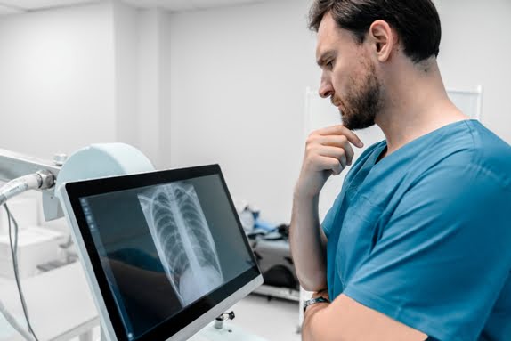
X Ray Both Leg AP Lat (₹ 1000)     Book An Appointment

Diagnopein X Ray Both Leg AP Lat Centre in Pune

Diagnopein X Ray Both Leg AP Lat Centre in Pune

An X-ray of Both Legs AP and Lateral (AP Lat) is a radiographic imaging technique used to capture detailed images of the bones in both legs, specifically focusing on the anterior-posterior (AP) and lateral views. This X-ray view is crucial for assessing the alignment, integrity, and any pathological changes in the bones of the legs, including the femur, tibia, fibula, and the joints of the knee and ankle. It is commonly used in the diagnosis and evaluation of bone fractures, deformities, infections, and various other conditions affecting the lower limbs.

X Ray Both Leg AP Lat Centre in Pune    Book Appointment

Purpose and Indications

1] Fractures and Trauma: One of the most common reasons for performing a bilateral leg X-ray is to detect fractures or other traumatic injuries to the bones. The AP and lateral views allow radiologists to examine fractures in the femur, tibia, fibula, and the bones of the ankle and knee. This is crucial for planning treatment, whether it be conservative management or surgical intervention.
2] Bone Deformities: X-rays help evaluate congenital or acquired deformities of the legs, such as bowlegs (genu varum), knock-knees (genu valgum), or abnormalities related to limb length discrepancies. These deformities can cause pain, instability, or difficulty walking.
3] Osteoarthritis and Joint Degeneration: Degenerative changes in the joints of the legs, particularly the knee, can be assessed with X-rays. The AP and lateral views can show joint space narrowing, bone spurs, and changes in bone density indicative of osteoarthritis or other degenerative conditions.
4] Infections and Bone Tumors: Conditions such as osteomyelitis (bone infection) or benign and malignant tumors affecting the bones of the legs can be detected. X-rays can show changes in the bone structure, including areas of bone destruction or abnormal growths.
5] Monitoring Post-Surgical Recovery: For patients who have undergone surgeries like fracture fixation, knee replacement, or leg lengthening, X-rays are used to monitor the healing process and ensure proper alignment and fixation of the bones.
6] Leg Length Discrepancy: In cases of leg length discrepancy, either due to congenital conditions or after fractures, X-rays provide measurements that can guide surgical correction or the use of orthotics.

For an accurate X-Ray Both Leg AP Lat in Pune, visit Diagnopein, the best digital X-ray test centre near me. This diagnostic test helps assess the bones, joints, and alignment of both legs, identifying conditions like fractures, arthritis, or deformities. Using cutting-edge digital X-ray technology, we provide high-resolution images with reduced radiation exposure. Our skilled radiologists analyze the results to assist in making precise treatment decisions. Whether you're experiencing pain or discomfort in your legs or need a routine evaluation, Diagnopein ensures quick, reliable, and professional X-ray services for your health and well-being.

Procedure and Technique

1] Patient Positioning:
A] AP View: The patient is asked to lie on their back on the X-ray table with both legs extended and slightly apart. The X-ray beam is directed from front to back, capturing both legs simultaneously to assess alignment and bone structures.
B] Lateral View: The patient is usually positioned on their side or, less commonly, lying on their back with both legs bent. The X-ray beam is directed from the side to provide a lateral view of the bones and joints of both legs.
2] X-ray Equipment Setup: The radiologic technologist ensures that the X-ray machine is correctly aligned to capture the required views. The exposure settings are adjusted based on the patient's size, the area of interest, and the clinical indications for the X-ray.
3] Imaging: Once the patient is in the correct position, the technologist will take the X-ray images. The patient may be asked to remain still and hold their breath briefly to reduce motion blur. Multiple images may be taken to ensure the necessary views are captured, especially if there is suspicion of fractures or deformities.
4] Post-Procedure: After the images are taken, the technologist will check the X-rays for clarity and accuracy. If necessary, additional images will be captured. The patient can then leave the imaging facility, and the images will be reviewed by a radiologist.
5] Image Interpretation: A radiologist will analyze the X-ray images for any signs of abnormalities. The findings will then be communicated to the referring physician, who will discuss the results with the patient and determine the appropriate course of action.

Diagnopein X Ray Both Leg AP Lat Treatement Cost     Book Appointment

Talk to Our Radiologist.

  I HereBy Consent to receive calls/messagess from Diagnopein Radiologist and its partners and override DND settings
faq image

Frequently Asked Questions

Yes, like all X-rays, the procedure involves exposure to a small amount of ionizing radiation. The radiation dose is low but should be minimized when possible.

It is used to diagnose conditions like bone fractures, deformities (e.g., bowlegs or knock-knees), osteoarthritis, and to assess the alignment and integrity of both legs.

The procedure is generally safe, though there is a slight risk due to radiation exposure. It is not recommended for pregnant women unless necessary.

No special preparation is needed. However, you may be asked to remove any metal objects (e.g., jewelry) that could interfere with the images.