What Is an X-Ray Femur AP/LAT?
This test captures X-ray images of the femur in two views:
-
AP (Anteroposterior) view
-
LAT (Lateral) view
These views provide a detailed assessment of the femur from different angles.
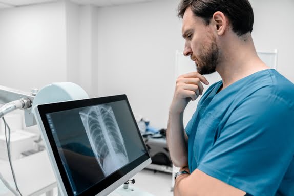
Digital X-rays ensure clear images with minimal radiation exposure.
Why Is X-Ray Femur AP/LAT Recommended?
Doctors recommend this test to evaluate femur-related conditions accurately.
It helps guide proper treatment and recovery planning.
Visit Now: https://www.diagnopein.com/digital-x-ray/Pune
Conditions Diagnosed Using Femur X-Ray
-
Femur fractures
-
Stress injuries
-
Bone infections (osteomyelitis)
-
Bone tumors or cysts
-
Post-surgical bone healing
Symptoms That May Require This Test
You may be advised to get this X-ray if you experience:
-
Severe thigh pain
-
Swelling after injury
-
Difficulty walking or standing
-
Pain after a fall or accident
-
Suspected bone injury
Early diagnosis helps prevent complications.
Procedure of X-Ray Femur AP/LAT
The procedure usually takes 10–15 minutes.
You will be positioned to allow imaging from the front and side of the thigh.
The radiology technician captures both views carefully.
The test is painless and completed quickly.
Preparation Before Femur X-Ray
Minimal preparation is required:
-
Remove metal objects from the area
-
Wear loose, comfortable clothing
-
Inform the technician if you are pregnant
No fasting or medication changes are needed.
Cost of X-Ray Femur AP/LAT in Pune
The cost of this test in Pune may vary based on:
-
Diagnostic center
-
Digital X-ray technology
-
Report turnaround time
Benefits of X-Ray Femur AP/LAT
-
Quick and accurate diagnosis
-
Digital imaging with low radiation
-
Helps detect fractures and bone changes
-
Affordable and widely available
-
Useful for injury and follow-up assessment
Who Should Book This Test?
This test is recommended for:
-
Patients with thigh injuries
-
Athletes with stress fractures
-
Elderly patients prone to fractures
-
Post-accident or trauma cases
Your orthopedic doctor will guide you on the need for this test.
Why Choose Diagnopen for Femur X-Ray in Pune?
-
Verified diagnostic centers
-
Modern digital X-ray equipment
-
Affordable and transparent pricing
-
Easy appointment booking
-
Fast and reliable reports
Diagnopen helps you find trusted imaging services easily.