


Diagnopein MRI Head – With Contrast Centre in Pune
Diagnopein MRI Head – With Contrast Centre in Pune
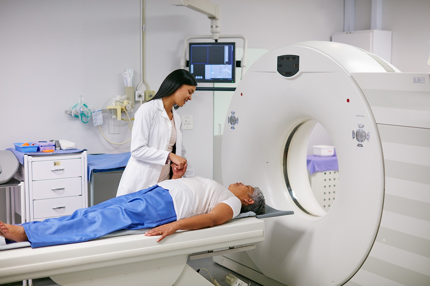
MRI (Magnetic Resonance Imaging) uses powerful magnetic fields and radio waves to generate detailed images of internal structures without the need for ionizing radiation. An MRI Head with Contrast specifically focuses on imaging the brain and its surrounding structures. The contrast agent, usually gadolinium-based, is injected into the patient’s bloodstream, enhancing the visibility of certain tissues, blood vessels, and abnormal growths.
The contrast agent works by altering the way MRI signals are absorbed and emitted by different tissues. Areas of the brain with abnormal blood flow, inflammation, or tumors will absorb the contrast agent differently than surrounding tissues, making them stand out more clearly on the scan.


