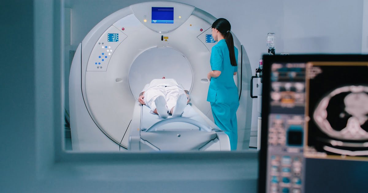
How Does a CT 3D Scan Work?
A CT 3D scan works by using multiple X-ray images taken from various angles. These images are processed by sophisticated computer software to generate a detailed three-dimensional model of the area being scanned. The process typically includes the following steps:
1. Patient Positioning: The patient is positioned on a special table that slides into the CT scanner. Depending on the body part being imaged, the patient may be asked to lie on their back, side, or stomach.
2. Image Acquisition: During the scan, the CT machine rotates around the patient, taking a series of X-ray images from different angles. The scanner captures these images in a series of thin slices, similar to how a loaf of bread is sliced.
3. 3D Reconstruction: The computer software reconstructs the multiple 2D slices into a 3D model. This process allows for detailed visualizations that can be rotated, zoomed in on, and viewed from multiple perspectives.
4. Contrast Dye: In many cases, a contrast dye may be injected into the patient’s veins or taken orally to enhance the images. The contrast helps highlight blood vessels, organs, and tissues, making abnormalities like tumors, fractures, or blockages easier to detect.
5. Analysis and Reporting: A radiologist examines the 3D images and prepares a report, which is shared with the referring physician. The images may also be used for planning surgeries or guiding interventions, such as biopsies or stent placements.
Why is a CT 3D Scan Important?
A 3D CT scan provides many advantages over traditional 2D CT imaging, especially in complex cases or when precise visualization of internal structures is required. Some of the key benefits include:
1. Detailed Visualization: The 3D model allows for better visualization of organs, bones, and tissues from any angle, which is useful for detecting small lesions, tumors, and other abnormalities that might not be as apparent in 2D images.
2. Surgical Planning: Surgeons often rely on 3D CT scans to plan complex surgeries. The 3D images help surgeons understand the relationship between different organs and tissues, allowing them to prepare for surgery with greater accuracy and precision.
3. Trauma Assessment: In cases of trauma, such as car accidents or falls, 3D CT scans can quickly and accurately assess injuries to bones, organs, and blood vessels, aiding in urgent medical decision-making.
4. Tumor Monitoring: For patients with cancer, 3D CT scans can be used to monitor the size and location of tumors over time. This allows for better tracking of treatment progress and adjustments to treatment plans if needed.
5. Vascular Imaging: The 3D CT scan is often used in CT angiography to visualize blood vessels in great detail. This is important for diagnosing conditions like aneurysms, arterial blockages, and vascular malformations, and it helps guide treatments like stent placement or embolization.
If you're looking for advanced imaging services, Diagnopein offers state-of-the-art 3D CT Scan in Pune. As a trusted CT scan centre in Pune, Diagnopein provides highly detailed, accurate 3D imaging that helps in precise diagnosis and treatment planning. The advanced technology ensures clear and accurate results, making it easier for doctors to detect issues that may be missed with traditional scans. With a focus on patient comfort and quick results, Diagnopein stands as a reliable choice for all your imaging needs in Pune.
Who Should Consider a CT 3D Scan?
A 3D CT scan is especially valuable for patients who require detailed, high-resolution imaging of internal structures. It is commonly used in the following situations:
1. Cancer Diagnosis and Treatment: Patients with tumors (e.g., lung, liver, or pancreatic cancer) can benefit from 3D CT scans to assess the size and spread of cancer. It helps in determining whether the tumor is operable and for planning radiation therapy.
2. Trauma Patients: Individuals who have been involved in traumatic accidents, such as car crashes, falls, or sports injuries, may need a 3D CT scan to evaluate fractures, internal bleeding, and damage to organs.
3. Surgical Planning: Surgeons use 3D CT scans to plan for complex surgeries, particularly in the abdominal area, brain, spine, or bones. The 3D images help identify the best approach and minimize the risk of complications.
4. Vascular Disorders: Patients with suspected vascular conditions, such as aneurysms or arterial blockages, may undergo a 3D CT angiography to visualize blood vessels and plan interventions like stent insertion or surgery.
5. Orthopedic Assessment: In orthopedics, 3D CT scans are used to evaluate bone fractures, joint abnormalities, or deformities. They provide a clearer understanding of complex bone injuries that may be missed on 2D scans.