Why X-Ray Right Foot AP/Oblique Is Important
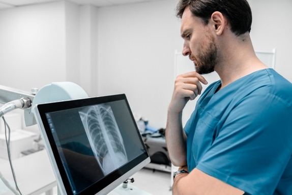
The X-Ray Right Foot AP/Oblique is important because it gives orthopaedic surgeons and physicians a clear visual understanding of bone structure and alignment. The AP view shows the foot in a straight-on position, whereas the oblique view helps highlight overlapping bones and joints, making hidden fractures or subtle deformities easier to identify.
Reasons Why It Is Important
-
Detects fractures – especially hairline or stress fractures that may not appear in a single view.
-
Assesses joint alignment – crucial for conditions such as arthritis, bunions, ligament injuries, and cartilage degeneration.
-
Evaluates foot deformities – helps diagnose flat feet, high arch, or alignment disorders.
-
Monitors post-treatment recovery – ideal for checking bone healing after surgery, plaster, or injury.
-
Supports early diagnosis – prevents complications by identifying bone infections or degenerative changes early.
At Diagnopein X Ray Centre Nashik, certified radiographers ensure optimal foot positioning to create accurate and diagnostically useful images.
Benefits of X-Ray Right Foot AP/Oblique
Getting a Right Foot X Ray AP/Oblique offers several clinical benefits, making it one of the most reliable diagnostic tools for foot-related issues.
Major Benefits
-
Quick & Non-Invasive – No injections or discomfort involved.
-
Accurate Structural Visualization – Provides a complete picture of bones, joints, and deformities.
-
Cost-Effective Imaging – More affordable compared to CT or MRI scans.
-
Immediate Reporting – At Diagnopein Nashik, digital X-Ray images are processed instantly.
-
Supports Orthopaedic Decisions – Helps doctors plan treatment for fractures, sports injuries, and chronic pain.
-
Minimal Radiation Exposure – Digital X-Ray equipment ensures safety.
This scan is especially helpful for athletes, elderly patients, accident victims, and individuals with chronic foot pain.
How the X-Ray Right Foot AP/Oblique Test Is Done
The procedure for an X-Ray Right Foot AP/Oblique at Diagnopein Nashik is simple, quick, and comfortable. Trained radiology experts guide the patient to ensure accurate positioning and minimal movement.
Step-by-Step Procedure
-
Positioning the Foot
The patient is asked to stand, sit, or lie down depending on comfort. The right foot is placed on the X-Ray plate in the AP position first.
-
Taking the AP View
The X-Ray beam is directed from front to back (anteroposterior) to capture the straight view of the foot.
-
Taking the Oblique View
The foot is rotated approximately 30–45 degrees to obtain a clear angled image showing overlapping bone structures.
-
Image Capture & Processing
Digital detectors instantly process the images for review. The radiologist examines the images for fractures, bone deformities, or alignment issues.
-
Report Generation
At Diagnopein Nashik, reports are usually available the same day with high-quality digital images.
Why Choose Diagnopein Nashik for X-Ray Right Foot AP/Oblique?
-
Advanced digital X-Ray systems for crystal-clear imaging
-
Experienced radiologists and technicians
-
Fast reporting with high diagnostic accuracy
-
Hygienic, patient-friendly environment
-
Affordable pricing for all X-Ray services
-
Centrally located reliable X-Ray Centre in Nashik
Keyword stuffing:Diagnopein Nashik X Ray, Best X Ray Centre Nashik, Right Foot X Ray Nashik, AP Oblique X Ray Foot.