X RAY RIGHT FEMUR AP/LAT (₹ 500)     Book An Appointment

Diagnopein X RAY RIGHT FEMUR AP/LAT Centre in Nashik

Diagnopein X RAY RIGHT FEMUR AP/LAT Centre in Nashik

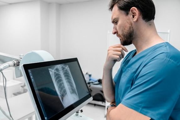
An X Ray Right Femur AP/LAT is a specialized imaging test that provides detailed views of the right femur from anteroposterior (AP) and lateral (LAT) perspectives. The femur, being the longest and strongest bone in the human body, is crucial for mobility and weight-bearing. Accurate imaging of this bone is essential for diagnosing fractures, infections, bone tumors, or alignment abnormalities.

X RAY RIGHT FEMUR AP/LAT Centre in Nashik    Book Appointment

Why X Ray Right Femur AP/LAT is Important

  1. Accurate Diagnosis: The AP and LAT views allow doctors to visualize the entire length of the right femur, ensuring no fractures or abnormalities are missed.

  2. Treatment Planning: Whether for orthopedic surgery, physiotherapy, or trauma care, thisX-rayprovides a complete assessment of bone structure and alignment.

  3. Early Detection: Conditions like bone infections, osteosarcoma, or osteoporosis-related fractures can be detected early, reducing complications.

  4. Monitoring Healing: For patients recovering from femur fractures or surgeries, X Ray Right Femur AP/LAT helps monitor healing progress accurately.


Benefits of X Ray Right Femur AP/LAT

  1. Non-invasive Procedure: No incisions or injections are required.

  2. Quick & Efficient: Typically, the procedure takes just a few minutes.

  3. Low Radiation Exposure: Modern digital X-rays minimize radiation dose while maintaining image quality.

  4. High Accuracy: Offers clear visualization of bone alignment, fractures, and lesions.

  5. Cost-Effective: Compared to other imaging methods like CT or MRI, X-rays are more affordable while delivering essential diagnostic information.

By choosing Diagnopein Nashik, patients benefit from expert radiologists who interpret X Ray Right Femur AP/LAT results with precision.


How X Ray Right Femur AP/LAT is Done

  1. Preparation: The patient may be asked to remove metal objects or clothing covering the thigh.

  2. Positioning for AP View: The patient lies on their back. The X-ray beam passes from front to back, capturing the femur in a straight-on view.

  3. Positioning for LAT View: The patient lies on their side. The X-ray beam passes from one side of the femur to the other, providing a side profile.

  4. Imaging: A digital X-ray system captures both AP and LAT images. The radiologist ensures the images are clear and cover the entire femur length.

  5. Analysis: The images are processed digitally and reviewed by a radiologist, who identifies fractures, lesions, or other abnormalities.


Why Choose Diagnopein Nashik for X Ray Right Femur AP/LAT

  1. Expert Radiologists: Skilled in interpreting femur X-rays with high precision.

  2. Advanced Equipment: Digital radiography ensures quick, high-resolution images.

  3. Patient Comfort: Safe, painless, and fast procedures.

  4. Comprehensive Reports: Detailed analysis provided for physicians and patients.

  5. Affordable Pricing: High-quality diagnostics at reasonable rates.

Patients in Nashik seeking reliable X Ray Right Femur AP/LAT imaging trust Diagnopein Centre for its commitment to accuracy, safety, and patient care.

Diagnopein X RAY RIGHT FEMUR AP/LAT Treatement Cost     Book Appointment

Talk to Our Radiologist.

  I HereBy Consent to receive calls/messagess from Diagnopein Radiologist and its partners and override DND settings