Why USG Fetal Echo? (Importance & Purpose)
A baby’s heart begins developing early in pregnancy, but some abnormalities may not be visible in a routine ultrasound. That’s why doctors recommend a dedicated fetal echocardiogram to assess cardiac health in detail.
You should consider USG Fetal Echo if:
-
You have abnormal NT scan, double marker, or anomaly scan results
-
You have diabetes, hypertension, thyroid disorders
-
There is a family history of congenital heart defects
-
You underwent IVF pregnancy
-
You are carrying twins / high-risk pregnancy
-
Exposure to certain medications or infections during pregnancy
The test helps detect:
-
Congenital Heart Disease (CHD)
-
Septal defects (ASD, VSD)
-
Valve abnormalities
-
Abnormal blood circulation
-
Heart rhythm disorders
-
Structural defects in chambers and arteries
Getting a USG Fetal Echo at Diagnopein Nashik ensures early diagnosis, which allows planning for timely medical intervention and improved baby outcomes.
Benefits of USG Fetal Echo at Diagnopein Nashik
Choosing Diagnopein Nashik for your USG Fetal Echo gives you several medical and diagnostic advantages:
Early Detection of Heart Problems
Detecting congenital heart conditions before birth helps parents prepare and doctors plan treatment steps.
Completely Safe & Radiation-Free Scan
The scan uses ultrasound waves, making it 100% safe for the mother and baby.
High-Resolution Fetal Imaging
Diagnopein uses advanced fetal ultrasound machines for superior image clarity.
Accurate Evaluation of Heart Function
The test analyses blood flow, heart rhythm, and structural alignment.
Essential for High-Risk Pregnancies
Helps in monitoring high-risk cases such as diabetes, thyroid disorders, and IVF pregnancy.
Expert Radiologists at Diagnopein Nashik
Highly trained fetal imaging specialists ensure precise and reliable reporting.
Improves Treatment Planning After Birth
Doctors can create a neonatal care plan in case of detected abnormalities.
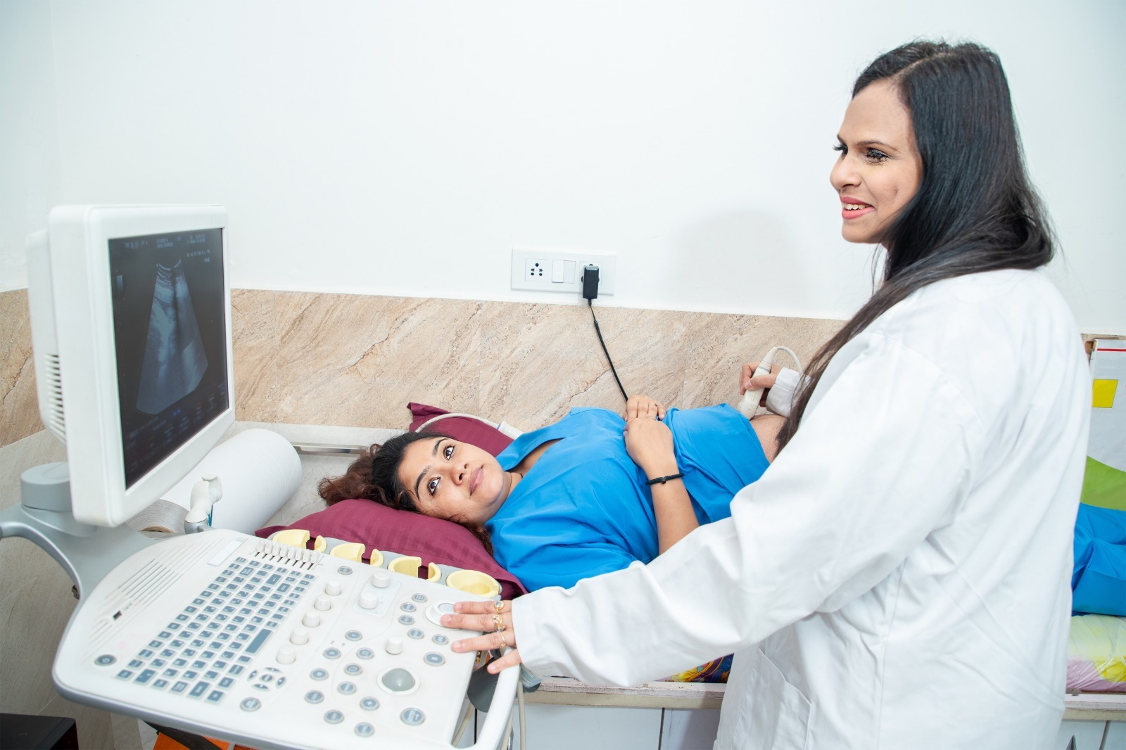
How USG Fetal Echo Is Done? – Procedure
The procedure is simple, painless, and takes around 25–40 minutes.
Step-By-Step Process at Diagnopein Nashik
-
Mother lies comfortably on the bed
-
Warm gel is applied on the abdomen
-
A high-frequency fetal echocardiography probe is used
-
The radiologist captures images of:
-
Heart chambers
-
Heart valves
-
Aorta & pulmonary artery
-
Blood flow patterns
-
Real-time Doppler imaging helps evaluate heart rhythm and flow
-
A detailed medical report is provided to your gynecologist
No fasting or preparation is required. You only need to carry your previous pregnancy reports.
Why Choose Diagnopein Nashik for USG Fetal Echo?
-
Advanced fetal imaging technology
-
Experienced radiologists
-
Accurate & detailed reporting
-
Comfortable scanning environment
-
Trusted by top gynecologists in Nashik
-
Affordable fetal echocardiography packages
-
High-risk pregnancy care support
Diagnopein Nashik is one of the most trusted centres for USG Fetal Echo, offering precision-based fetal heart evaluation.