Importance of CT RT Wrist Joint Scan
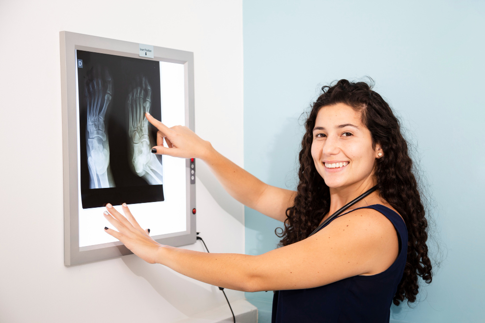
The wrist is one of the most complex joints in the human body, comprising tiny bones and ligaments that control daily movement. A CT RT Wrist Joint scan helps in detecting conditions like stress fractures, dislocations, cysts, bone tumors, or degenerative changes in wrist cartilage.
Orthopedic specialists often recommend this scan when patients experience chronic wrist pain, swelling, or mobility limitations not clearly explained by preliminary tests. CT imaging provides a multi-angle, cross-sectional view that assists in evaluating:
-
Fine bone details and subtle cracks.
-
The extent of ligament or tendon injury.
-
Healing progress after a fracture or surgery.
-
Bone density and microstructure.
At Diagnopein Nashik, performing this CT scan aids in timely detection of problems, supports treatment precision, and improves recovery outcomes through evidence-based imaging.
Benefits of CT RT Wrist Joint Imaging
The CT Right Wrist Joint scan offers several advantages compared to conventional imaging methods.
-
High clarity and accuracy: CT technology captures detailed cross-sectional images that highlight even minute abnormalities.
-
Quick and painless: The entire scan takes only a few minutes and does not require any special preparation.
-
3D reconstruction: Radiologists at Diagnopein Nashik can create digital 3D models for better visualization of wrist anatomy.
-
Effective for trauma cases: Ideal for analyzing complex fractures and confirming bone alignment post-plaster or surgery.
-
Low radiation dose: Modern CT scanners use optimized settings ensuring patient safety while maintaining image quality.
Because wrist movement affects comfort and recovery, accurate diagnosis through this scan helps plan physiotherapy, surgical repair, or medical management appropriately.
Procedure, Placement, and Parameters
Preparation and Placement
The CT RT Wrist Joint scan is performed with the patient seated or lying on a CT table. The right wrist is positioned carefully inside the scanner with supportive cushions to avoid movement. Patients are asked to remove any metallic objects such as watches or bangles.
Procedure Steps
-
The technologist positions the wrist correctly in the scanning field.
-
The CT machine rotates around the wrist, capturing multiple thin-slice images.
-
Advanced software reconstructs these slices into a clear, 3D, high-resolution image.
-
The radiologist then analyzes the images for structural integrity and alignment.
Scan Parameters
Typical technical parameters include:
-
Scan type: Axial and coronal reconstruction.
-
Slice thickness: 0.5–1.25 mm.
-
Voltage (kVp): 120.
-
Exposure time: Few seconds.
-
Contrast usage: Optional, depending on soft tissue evaluation needs.
After the Test
No recovery time is required after the procedure. Patients can continue daily activities immediately. The scan images and radiology report are available the same day at Diagnopein Nashik, ensuring faster clinical consultation.
Why Choose Diagnopein Nashik for CT Wrist Joint Scan?
Diagnopein offers cutting-edge imaging technology operated by certified radiologists and technologists. The center emphasizes:
-
Fast and accurate reporting.
-
Affordable pricing with transparent billing.
-
Hygienic and patient-friendly environment.
-
Personalized support for each patient.
Choosing Diagnopein Nashik ensures comfort, reliability, and diagnostic accuracy that helps your doctor make the best treatment decision.