Importance of CT Pelvis with 3D Placement
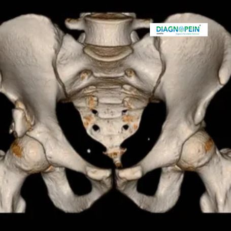
The CT Pelvis with 3D placement offers a complete view of internal structures, making it highly reliable for identifying fractures, tumors, abscesses, or infections. This scan plays an essential role in evaluating trauma cases and pre-surgical planning, especially in orthopedic and urological conditions.
Unlike traditional X-rays, a 3D CT pelvis provides volumetric data that allows radiologists to assess bone alignment, joint spaces, and soft-tissue involvements without invasive procedures. In female patients, it helps assess uterine and ovarian disorders, while in males, it aids in prostate evaluations.
By combining accuracy, speed, and versatility, Diagnopein Nashik’s CT Pelvis with 3D scan ensures early detection and proper treatment guidance.
Benefits of CT Pelvis 3D Scan
The 3D CT pelvis scan provides outstanding diagnostic benefits that improve healthcare outcomes:
-
Detailed visualization: Offers highly detailed 3D images for a comprehensive view of pelvic anatomy.
-
Accurate diagnosis: Detects fractures, infections, and abnormalities with superior precision.
-
Non-invasive procedure: Provides deep internal insights without surgery or discomfort.
-
Quick results: Produces fast and accurate results suitable for emergency and routine assessments.
-
Enhanced surgical planning: 3D models help surgeons plan interventions more accurately.
Patients visiting Diagnopein Nashik can expect expert radiologists, modern CT technology, and precise reporting for confident diagnosis.
CT Pelvis with 3D Testing Procedure and Parameters
The CT Pelvis with 3D test at Diagnopein Nashik follows a fast and patient-friendly procedure:
-
The patient lies comfortably on a motorized CT table.
-
Contrast dye may be administered intravenously for better visualization of internal organs and blood vessels.
-
The scanner rotates around the pelvic area to capture multiple X-ray images.
-
The computer reconstructs these slices into detailed 3D images for accurate study and diagnosis.
The typical scan duration is 10–20 minutes depending on the clinical requirement. Patients are advised to avoid metal objects and fasting may be required if contrast material is used.