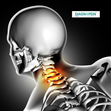
Importance and Placement of CT Mandible with 3D
The CT Mandible 3D scan plays a crucial role in diagnosing mandibular disorders and pre-surgical planning. It is recommended for cases involving trauma, dislocation, dental implant evaluation, cysts, and tumor detection.
Placement refers to how the patient is positioned during the scan. The patient lies supine on the CT table with the head stabilized to avoid movement. Proper placement ensures precise imaging of the mandible region and prevents distortion in 3D reconstruction.
In Nashik, Diagnopein Diagnostic Center provides accurate CT Mandible imaging under expert supervision, ensuring comfort and safety for every patient. This test helps specialists understand bone dimensions and alignments critical for surgical or dental correction procedures.
Benefits of CT Mandible with 3D Scan
A CT Mandible with 3D scan offers several advantages over conventional X-rays or 2D imaging. Some key benefits include:
-
High-resolution 3D visualization of jawbone, teeth roots, and surrounding tissues.
-
Accurate localization of infections, fractures, or foreign bodies in the mandible.
-
Improved surgical planning, particularly for dental implants, trauma reconstruction, and oral tumor removal.
-
Quick and painless procedure with instant digital image generation.
-
Reduced errors in facial or dental measurements due to 3D precision.
Patients in Nashik prefer getting this scan at Diagnopein because of advanced CT systems, experienced radiologists, and reliable diagnostic reports.
Procedure of CT Mandible with 3D
The CT Mandible with 3D test is quick, simple, and performed without discomfort. Below are the typical steps:
-
The patient removes metallic jewelry or dental appliances to avoid image artifacts.
-
The radiology technician positions the patient comfortably on the CT scanning bed.
-
A thin X-ray beam rotates around the head to capture multiple cross-sectional images of the jawbone.
-
The computer processes these slices to form a detailed 3D reconstruction.
-
The entire scan usually completes within 5 to 10 minutes.
-
Images are reviewed by radiologists before generating a diagnostic report.
For some evaluations, a minor contrast material may be used to enhance tissue visibility, depending on the doctor’s suggestion.
CT Mandible Scan Parameters
Typical technical parameters used for CT Mandible with 3D in Nashik include:
-
Scan Type: Multi-slice helical CT
-
Slice Thickness: 0.5–1 mm
-
Reconstruction Interval: 0.3–0.5 mm
-
Field of View (FOV): 120–180 mm
-
Radiation Dose: Low-dose adaptive protocol
-
Reconstruction Algorithm: Bone (High-resolution) and Soft Tissue
At Diagnopein Nashik, these parameters are tailored to provide maximum diagnostic detail with minimal radiation exposure.
Why Choose Diagnopein Nashik for CT Mandible 3D
Diagnopein Nashik is one of the trusted names for CT Mandible with 3D imaging because of its advanced diagnostic technology and patient-friendly service. The center ensures precision through fully digital CT systems, expert radiologists, and quick result delivery. Patients benefit from clear, accurate 3D jaw visualization helpful for surgical and dental treatment planning.
Whether it’s for trauma, dental implants, or jaw pathology, Diagnopein Nashik provides reliable imaging results to support accurate medical decisions.