3D SONOGRAPHY IMAGE (₹ 1000)     Book An Appointment

Diagnopein 3D SONOGRAPHY IMAGE Centre in Nashik

Diagnopein 3D SONOGRAPHY IMAGE Centre in Nashik

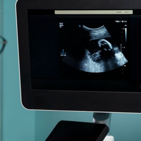
3D Sonography Image, also called 3D ultrasound, is an advanced medical imaging technique that creates three-dimensional images of the body's internal structures. Unlike traditional 2D ultrasound that produces flat images, 3D sonography offers volumetric views of tissues and organs, providing more detailed visualization. This technology is commonly used in obstetrics, gynecology, cardiology, and musculoskeletal imaging to aid in precise diagnosis and treatment planning. The ability to view the anatomy in three dimensions helps physicians understand complex conditions better and improves patient care.

3D SONOGRAPHY IMAGE Centre in Nashik    Book Appointment

Importance of 3D Sonography Image

  1. Why 3D Sonography Matters
    3D ultrasound imaging is important because it enhances diagnostic accuracy by showing more realistic views of the area being examined. It allows visualization of fetal features in prenatal care like facial structure and limb positioning, helping detect abnormalities early. In other fields such as cardiology, 3D sonography visualizes heart structures dynamically, aiding in the assessment of heart function and defects. The volumetric data provided by 3D sonography also supports better surgical planning and monitoring treatment efficacy, making it a critical tool in modern medical practice.


Benefits of 3D Sonography Image

  1. Advantages You Gain
    Choosing 3D sonography at Diagnopein offers numerous benefits:

  2. Enhanced Image Clarity: Three-dimensional imaging provides clear, lifelike images, improving diagnostic confidence.

  3. Non-Invasive and Safe: Uses sound waves without radiation, making it safe for all patients, including expectant mothers.

  4. Real-Time Visualization: Enables real-time monitoring of organs and fetus movements.

  5. Comprehensive Analysis: Volumetric data allows examination from multiple angles and planes for thorough evaluation.

  6. Early Detection: Helps detect anomalies or issues sooner, leading to better outcomes.


How to Do 3D Sonography Testing

  1. Testing Procedure and Parameters

    The 3D sonography test is straightforward and painless. A trained sonographer applies a water-based gel on the skin over the area to be examined to ensure good contact with the transducer. The transducer emits sound waves, capturing multiple 2D images from different angles which are then digitally reconstructed into a 3D volume. Testing typically takes 15 to 30 minutes depending on the region scanned. Important parameters analyzed include organ shape, volume, surface structure, and internal tissue details. Patients are advised to follow any pre-test instructions given by their healthcare provider for best results.


Why Choose Diagnopein in Nashik for 3D Sonography Image?

  • State-of-the-art imaging equipment providing high-resolution 3D images.

  • Expert radiologists and sonographers with extensive experience.

  • Comfortable and patient-friendly environment for all diagnostic procedures.

  • Timely and accurate reporting to facilitate prompt medical decisions.

  • Affordable pricing with advanced technology services accessible in Nashik.

Diagnopein 3D SONOGRAPHY IMAGE Treatement Cost     Book Appointment

Talk to Our Radiologist.

  I HereBy Consent to receive calls/messagess from Diagnopein Radiologist and its partners and override DND settings
faq image

Frequently Asked Questions

While 3D ultrasound provides highly detailed images, it is not always able to detect all types of fetal abnormalities. Some conditions may require additional imaging tests, such as 2D ultrasound, MRI, or amniocentesis, to confirm a diagnosis. However, 3D ultrasound helps identify physical abnormalities like facial clefts, spina bifida, or heart defects.

A 3D ultrasound generally takes 30 to 45 minutes, depending on the complexity of the imaging and the area being examined. The procedure may take longer if multiple angles or detailed views are required.

Yes, 3D sonography is completely safe for both the mother and the baby. It uses sound waves, not radiation, and is a non-invasive procedure. It is generally considered as safe as traditional 2D ultrasound.

3D ultrasound is usually performed after 12 weeks of pregnancy. The best time to get clear images is typically between 26 and 30 weeks, when the baby has enough fat for clearer visualizations, but before the baby’s position becomes too cramped.