The HPE Breast Sample (Histopathological Examination) is a crucial test used to study breast tissue under a microscope. It helps in diagnosing breast cancer, benign lumps


The HPE Breast Sample test, or Histopathological Examination of Breast Tissue, is one of the most definitive diagnostic tools for identifying breast cancer and other breast disorders. The test involves microscopic analysis of tissue collected from a breast lump, biopsy, or surgical specimen, helping pathologists detect abnormal cell growth, inflammation, or tumors.
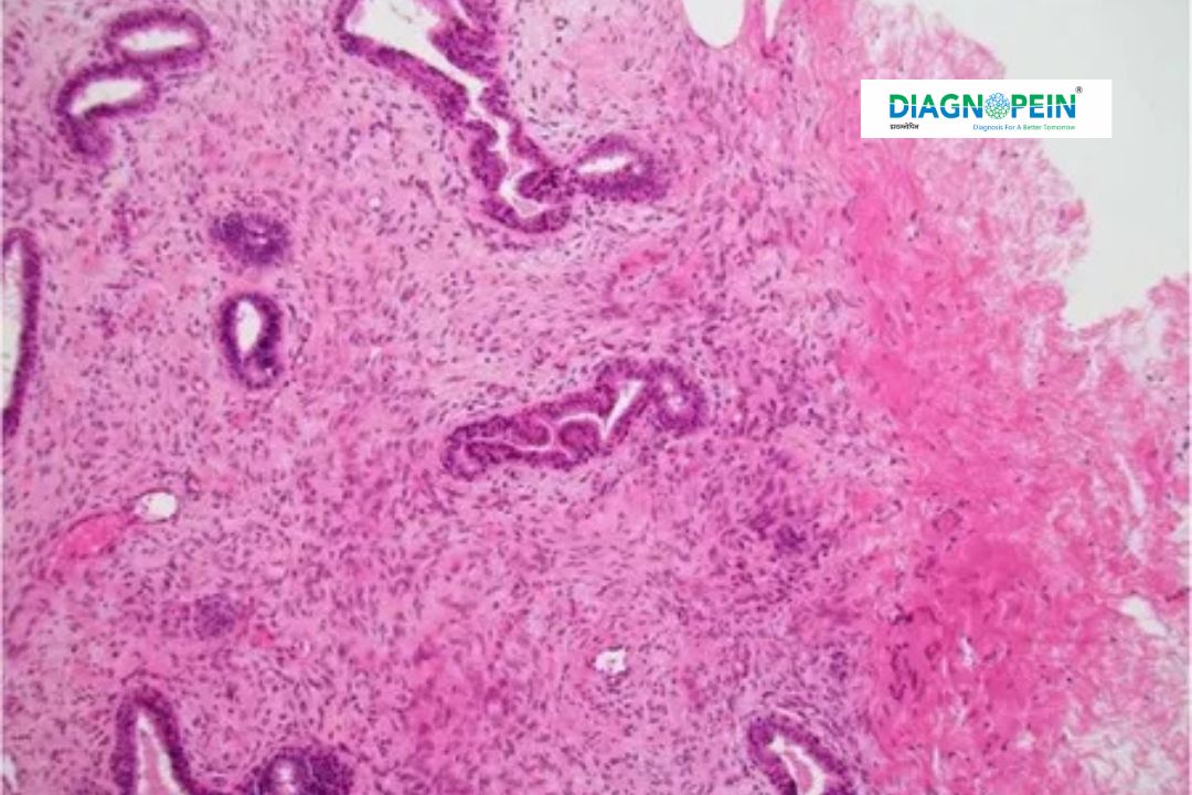
Histopathology remains the gold standard for diagnosing breast diseases because it provides direct insights into tissue architecture and cellular characteristics. Accurate diagnosis through HPE Breast Sample testing allows doctors to determine the exact stage and type of disease, enabling suitable clinical management.
Breast diseases, including infections, fibrocystic changes, and breast cancer, share similar symptoms like lumps or pain, making histopathological confirmation essential. The HPE breast test provides the final confirmation about whether a lump is benign or malignant. It also helps in differentiating conditions such as fibroadenoma, ductal carcinoma, and lobular carcinoma.
Early detection and timely diagnosis are life-saving. The HPE Breast Sample test helps oncologists determine treatment protocols, such as surgery, chemotherapy, or hormonal therapy. This makes it a vital part of every breast cancer diagnostic process.
Provides accurate and detailed diagnosis of breast tissue abnormalities.
Helps clinicians distinguish between benign lesions and cancerous tumors.
Enables early detection, ensuring timely intervention and treatment.
Assists in staging of breast cancer, guiding appropriate treatment plans.
Offers valuable information on tumor margins after surgery.
Cost-effective and reliable diagnostic procedure compared to imaging alone.
For women above 40 or those with a family history of breast cancer, undergoing an HPE breast test after a biopsy or surgery offers peace of mind and clarity about their condition.
Sample Collection – A small portion of breast tissue is obtained through needle biopsy, lumpectomy, or mastectomy.
Fixation – The sample is preserved in formalin to prevent decomposition.
Processing & Sectioning – The tissue is embedded in paraffin, sliced into thin sections, and placed on glass slides.
Staining – Special dyes such as Hematoxylin and Eosin make cell structures visible under a microscope.
Microscopic Analysis – A pathologist examines the slide to detect abnormal cells, tumor type, and stage.
Report Generation – A detailed HPE report is prepared, providing diagnostic findings and clinical recommendations.
The entire process generally takes 3–5 days, depending on laboratory workload and sample complexity.
Cell structure and morphology
Presence of atypical or malignant cells
Mitotic activity count (cell division rate)
Tumor infiltration and margin status
Inflammatory or necrotic tissue changes
Histological tumor grade and pattern
These parameters ensure a definitive diagnosis and guide oncologists in personalizing each patient’s treatment journey.
At Diagnopein, we prioritize precision, safety, and timely results for every HPE Breast Sample test.
Experienced pathologists specializing in breast histopathology.
Advanced laboratory equipment for accurate analysis.
100% confidentiality and online report access.
Affordable HPE breast test price with quick turnaround time.
Personalized patient support and consultation services.
Choose Diagnopein for dependable diagnostic solutions and step forward confidently in your healthcare journey.

While the biopsy is generally safe, there are minor risks involved, such as bleeding, infection, and bruising at the biopsy site. Discuss any concerns with your healthcare provider before the procedure.
A positive result indicates the presence of cancerous cells in the tissue sample. This requires further evaluation and discussions regarding treatment options with healthcare providers.
The breast tissue sample can be collected through various biopsy methods, including Fine Needle Aspiration (FNA), Core Needle Biopsy, Excisional Biopsy, or Incisional Biopsy. The method used depends on the size and location of the suspicious area.
The level of discomfort varies by the type of biopsy performed. Fine Needle Aspiration typically involves minimal discomfort, while excisional biopsies may require anesthesia, resulting in a pain-free experience during the procedure.


