Expecting a baby is one of the most exciting times in a parent's life, but it also comes with the responsibility of ensuring the health and well-being of both the mother and the baby. One essential procedure that many expectant parents may be unfamiliar with is fetal echocardiography. This specialized ultrasound helps monitor the health of your baby’s heart during pregnancy, and it plays a crucial role in detecting potential heart conditions before birth.
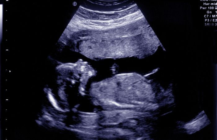
Fetal echocardiography, often referred to as a fetal echo, is a non-invasive ultrasound exam that uses sound waves to create detailed images of the baby's heart while still in the womb. It allows doctors to assess the structure and function of the heart, detecting any abnormalities such as congenital heart defects, which are among the most common birth defects.
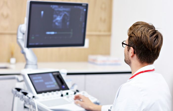
Unlike routine ultrasounds, which mainly focus on the baby’s overall development and anatomy, fetal echocardiography is specifically designed to examine the heart. The procedure involves applying a special gel to the abdomen of the mother, followed by a handheld device called a transducer, which emits sound waves to capture real-time images of the fetal heart.
Fetal echocardiography is critical for ensuring the baby’s heart health, particularly in cases where there is a concern about a potential heart problem. Around 1 in 100 babies is born with a congenital heart defect, and some may not show symptoms until later in life. Fetal echocardiography helps identify heart conditions early, allowing for timely interventions and treatment options if needed.
Early detection of heart problems through fetal echocardiography can help prepare parents for what to expect after birth, ensuring that the necessary medical team is ready to assist in the care of the baby immediately after delivery. It can also reduce the risk of complications by detecting abnormalities that may require surgery or other treatments soon after birth.
Fetal echocardiography is not routinely performed for every pregnancy. Instead, it is generally recommended when there are certain risk factors or concerns about the baby's heart. These may include:
1] Family history of heart disease: If a parent or close relative has a history of congenital heart disease, there may be a higher risk of the baby having a similar condition.
2] Abnormal findings in a routine ultrasound: If a routine ultrasound reveals potential heart abnormalities, a fetal echocardiogram may be advised for a more detailed examination.
3] Maternal health conditions: Conditions like diabetes, lupus, or certain infections during pregnancy can increase the risk of heart defects in the baby.
4] Genetic conditions: If there is a suspicion of a genetic disorder that may affect the baby’s heart, fetal echocardiography can help confirm or rule out heart defects.
5] Increased nuchal translucency: A thickened nuchal fold seen in the routine ultrasound may suggest an increased risk of heart problems.
If any of these factors apply to you, your healthcare provider may recommend a fetal echo to ensure your baby’s heart health is properly assessed. In most cases, this procedure is done between 18 and 22 weeks of pregnancy, although it can be performed later if necessary.

Fetal echocardiography is a straightforward and non-invasive procedure. Here’s what you can expect during the test:
No special preparation is usually required for the procedure, though your healthcare provider may ask you to drink water before the exam to help improve the quality of the images. It's a good idea to wear loose, comfortable clothing for the session.
The procedure itself involves applying a gel to the abdomen, which helps the ultrasound transducer make contact with the skin and send sound waves to create the images. The technician will move the transducer across the abdomen to get different views of the baby’s heart. You may be asked to change positions to help get the best possible images.
Fetal echocardiography typically takes about 30 minutes to an hour, depending on the baby’s position and the clarity of the images. In some cases, the procedure may take longer if additional views or images are needed.
After the procedure, the images are analyzed by a specialist who will interpret the results. If any abnormalities are detected, further tests or referrals to a pediatric cardiologist may be necessary to confirm the diagnosis and determine the best course of action.
The cost of fetal echocardiography can vary depending on the location, healthcare facility, and the complexity of the procedure. On average, the cost for fetal echocardiography at Diagnopein is Rs. 3000/-. This includes the full evaluation and interpretation of the images by qualified specialists to ensure the best care for both you and your baby.
If you're looking for a sonography centre near me in cities like Bhopal, Mumbai, Pune, Nagpur, or Delhi, Diagnopein is a trusted name offering high-quality fetal echocardiography services. We are committed to providing accurate results and comprehensive care to ensure the health of your baby.
Diagnopein is a leading healthcare provider with a reputation for excellence in diagnostic services. If you're expecting and need a sonography centre near me for fetal echocardiography, Diagnopein offers:
Our team of experienced sonographers and specialists is dedicated to providing accurate, high-quality imaging to ensure the best care for your baby. Our staff is highly trained to perform fetal echocardiography with the utmost precision and care.
At Diagnopein, we use the latest ultrasound technology to provide clear, detailed images of your baby's heart. Our equipment is regularly updated to ensure the highest standard of care.
With locations in Bhopal, Mumbai, Pune, Nagpur, and Delhi, we make it easy for expecting parents to find a convenient sonography centre near me for fetal echocardiography and other essential pregnancy-related tests.
Our services are priced competitively to make prenatal care accessible. With a cost of Rs. 3000/- for fetal echocardiography, you can ensure that both you and your baby receive the best possible care without breaking the bank.