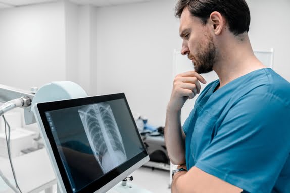
Why is an X-Ray Skull AP Performed?
1] Fractures: This is one of the primary reasons for a skull X-ray. Trauma to the head can lead to fractures of the cranial bones, and the AP view helps identify fractures in the frontal, parietal, temporal, and occipital bones.
2] Sinus Issues: The AP view provides a clear image of the sinuses (frontal, maxillary, ethmoid, and sphenoid), allowing the radiologist to detect conditions like sinusitis or fluid buildup in the sinuses.
3] Tumors and Lesions: Abnormal growths such as tumors, cysts, or abscesses in the skull or sinuses can be identified. A skull X-ray can help detect any irregularities or mass effect caused by these growths.
4] Congenital Abnormalities: The AP view can reveal craniosynostosis (early closure of skull sutures) and other birth defects that may cause abnormalities in skull shape and structure.
5] Infections: Infections affecting the skull, such as osteomyelitis (bone infection), can cause visible changes in bone structure. An X-ray may help confirm such conditions.
For an X-Ray Skull AP in Pune, Diagnopein is the best digital X-ray centre near me. This test provides detailed images of the skull, helping in the diagnosis of fractures, infections, tumors, or abnormalities in the bone structure. Using advanced digital X-ray technology, we ensure clear, high-resolution images with reduced radiation exposure. The X-ray results are quickly analyzed by our expert radiologists to offer accurate diagnostic reports. Whether it's for trauma, routine check-ups, or medical evaluations, Diagnopein offers efficient and reliable skull X-ray services, ensuring you receive the best possible care in Pune.
How the Procedure is Performed ?
To perform the AP skull X-ray, the patient typically sits or stands with the head facing directly forward. The following steps are involved:
1] Positioning: The patient is positioned with their forehead against the X-ray machine. The chin is slightly tilted to ensure that the skull is aligned properly.
2] Image Capture: The X-ray machine sends a small amount of radiation through the skull, and the detector captures the image from front to back.
3] Multiple Images: Depending on the clinical situation, multiple images may be taken from different angles, such as lateral or oblique views, to get a comprehensive understanding of the skull's condition.


