USG SINGLE BREAST (₹ 2500)     Book An Appointment

Diagnopein USG SINGLE BREAST Centre in Pune
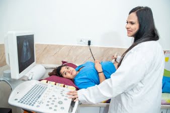
Diagnopein USG SINGLE BREAST Centre in Pune

USG Single Breast, also known as breast ultrasound, is a non-invasive imaging technique used to examine breast tissue. It helps in the evaluation of any abnormalities or changes in the breast, such as lumps, cysts, or other conditions. Unlike a mammogram, which uses X-rays, a breast ultrasound uses sound waves to create detailed images of the breast's internal structures.

USG Single Breast in Pune is a crucial diagnostic tool to assess breast health and detect any abnormalities. At Diagnopein, the best sonography centre in Pune, we offer high-quality ultrasound services for detailed evaluation of the breast tissue. Our expert technicians ensure precise imaging for early detection of conditions like fibroadenomas or tumors. Trust Diagnopein for accurate, reliable results and comprehensive care at the best sonography centre in Pune, committed to your health and well-being.

USG SINGLE BREAST Centre in Pune    Book Appointment

Why is a Single Breast Ultrasound Important?

A single breast ultrasound is typically performed to investigate specific symptoms or concerns related to one breast, such as:
1. Evaluation of Lumps: If a lump is detected in one breast during self-examination, a clinical exam, or a mammogram, a breast ultrasound can help determine the nature of the lump (e.g., cystic or solid), which helps in differentiating between benign conditions and potential tumors.
2. Assessment of Breast Pain: Breast pain (mastalgia) localized to one breast can be evaluated to determine if it's related to cysts, fibroadenomas, or other benign breast conditions.
3. Guiding Biopsy: If a suspicious mass is found, a breast ultrasound may be used to guide a biopsy needle to the exact location for sampling tissue for further examination.
4. Evaluation of Cysts: Ultrasound can clearly distinguish between a solid mass (which may require further investigation) and a fluid-filled cyst (which is often benign). It can also monitor the size and changes in existing cysts.
5. Examination of Dense Breast Tissue: For women with dense breast tissue, which can make it more difficult to detect abnormalities through mammograms, an ultrasound provides a clearer picture of any potential issues.

Who Should Consider a Single Breast Ultrasound?

A single breast ultrasound is typically recommended for:
1. Women with a Lump in One Breast: If you notice a lump or mass in one breast, ultrasound is often used to evaluate its nature (cystic or solid) and to check for any potential risk factors.
2. Women with Localized Breast Pain: If breast pain is concentrated in one area, an ultrasound can help identify the cause, whether it's related to benign conditions like cysts or fibroadenomas.
3. Follow-Up After a Mammogram: If a mammogram shows an abnormality in one breast, an ultrasound may be used as a follow-up tool to gather more information and assess the area in more detail.
4. Women with Dense Breast Tissue: Women with dense breasts (which makes mammogram results harder to interpret) often benefit from an ultrasound to help detect any hidden abnormalities.

Diagnopein USG SINGLE BREAST Treatement Cost     Book Appointment

Talk to Our Radiologist.

  I HereBy Consent to receive calls/messagess from Diagnopein Radiologist and its partners and override DND settings
faq image

Frequently Asked Questions

No, a breast ultrasound is painless. You may feel some pressure or slight discomfort when the transducer is moved over the breast, but it is not painful.

Yes, a breast ultrasound is completely safe. Unlike mammograms, it does not use radiation, making it ideal for women who need repeated screenings or are pregnant.

A breast ultrasound can help detect abnormalities such as lumps or cysts, but it cannot definitively diagnose breast cancer. If a suspicious area is found, further tests such as a biopsy or mammogram may be necessary to confirm the diagnosis.

The results of a breast ultrasound are usually available within 1-2 days after the procedure. The radiologist will analyze the images and send the report to your doctor, who will discuss the findings with you.