Why Would You Need an MRI Brain Cranial Nerve Protocol?
An MRI with cranial nerve protocol is typically ordered when there are symptoms that suggest issues with the brain or cranial nerves. Common reasons for undergoing this MRI scan include:
1] Neurological Symptoms: If a person is experiencing unexplained headaches, dizziness, facial weakness, numbness, double vision, or difficulty swallowing, an MRI may be recommended to determine if any cranial nerves are being affected.
2] Tumor Detection: MRI with cranial nerve protocol is commonly used to detect brain tumors or abnormal growths that might put pressure on the cranial nerves. Conditions such as acoustic neuromas, pituitary tumors, or meningiomas are best detected using MRI.
3] Cranial Nerve Lesions: In cases of trauma, infections, or inflammation, the cranial nerves may be damaged. An MRI can help identify any lesions or abnormalities in the nerve pathways.
4] Multiple Sclerosis (MS): MS can cause lesions in the brain that affect the cranial nerves. A cranial nerve MRI protocol is used to assess any damage or plaques in the brain associated with MS.
5] Vascular Conditions: Strokes, aneurysms, or abnormal blood vessels can impact the cranial nerves. The MRI scan can detect such vascular issues that may be impairing normal nerve function.
6] Infection or Inflammation: Conditions like meningitis or encephalitis can cause swelling in the brain and affect the cranial nerves. MRI helps in detecting any inflammation in the brain tissues or along the nerve pathways.
MRI Brain Cranial Nerve Protocol Cost in Pune
The cost of an MRI Brain Cranial Nerve Protocol in Pune is typically around Rs. 3900. This specialized MRI scan is used to evaluate the brain and cranial nerves, helping to diagnose conditions like tumors, nerve compression, multiple sclerosis, or neurological disorders. The price may vary depending on the diagnostic center, the MRI technology used, and additional services provided. For accurate pricing and availability, it’s recommended to consult local MRI centers or hospitals in Pune.
How is an MRI Brain Cranial Nerve Protocol Performed?
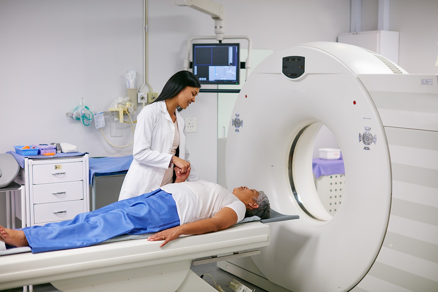
The MRI procedure itself is non-invasive and typically lasts between 30 to 60 minutes. Here's a step-by-step overview of what to expect during an MRI brain cranial nerve protocol:
1] Preparation: No special preparation is typically required, but you may be asked to remove any jewelry, hairpins, or metal items that can interfere with the magnetic field. If you're having contrast dye administered (which is sometimes used to improve image clarity), you may be asked to fast for a few hours before the scan.
2] Positioning: During the scan, you will lie on a table that slides into the MRI machine. You will be positioned to ensure that your head is in the proper alignment for imaging the brain and cranial nerves. A head coil may be used to stabilize your head during the procedure.
3] The Scan: The MRI machine uses magnetic fields and radio waves to create detailed images. The procedure is painless, though you may hear a loud tapping or knocking sound during the scan. You will be asked to remain as still as possible to ensure clear images are obtained.
4] Contrast Dye: In some cases, contrast dye (gadolinium-based) may be injected into your vein during the scan. This helps improve the clarity of the images, especially when examining vascular structures or tumors.
5] Post-scan: Once the MRI scan is completed, you can resume your normal activities. If contrast dye was used, you may be advised to drink plenty of fluids to flush it out of your system.