Why is MRI Angiography with Contrast Performed?
1] Detecting Aneurysms: One of the primary uses of MRA is to identify aneurysms—bulging, weakened areas in the walls of blood vessels that can rupture and cause life-threatening bleeding. MRI Angiography can detect aneurysms in various areas, including the brain (cerebral aneurysms), heart (aortic aneurysms), and peripheral arteries.
2] Assessing Arterial Blockages and Stenosis: MRA is commonly used to evaluate arteries for blockages, narrowing (stenosis), or other forms of obstruction, particularly in the carotid, renal, or coronary arteries. This is critical for diagnosing conditions like atherosclerosis (hardening of the arteries) or peripheral artery disease (PAD).
3] Evaluating Blood Flow: MRI Angiography can assess the flow of blood through the vascular system, helping doctors understand how well blood is reaching vital organs and tissues. It is particularly useful in evaluating conditions that affect circulation, such as deep vein thrombosis (DVT) or venous insufficiency.
4] Pre-surgical Planning: MRA is often used before vascular surgery or endovascular procedures, such as stent placements or bypass surgeries. It provides detailed images of the blood vessels to help surgeons plan their procedures more effectively.
5] Diagnosing Vascular Malformations: MRA can be used to identify vascular malformations or abnormal connections between arteries and veins. These conditions, which include arteriovenous malformations (AVMs) and fistulas, can cause a range of symptoms depending on their location.
6] Monitoring Known Vascular Conditions: Patients with a history of vascular conditions like aneurysms, arteriosclerosis, or blood clots may undergo periodic MRI Angiography to monitor their condition and assess whether it is progressing or improving.
How Does MRI Angiography with Contrast Work?
1] Preparation: Before the scan, the patient may be asked to fast for a few hours, especially if the scan is being performed to evaluate blood vessels in the abdominal or chest regions. The patient should also remove any metallic objects, such as jewelry, hearing aids, or dentures, as metal can interfere with the MRI machine.
2] Contrast Injection: A contrast agent (gadolinium) is injected into a vein, typically in the arm, to enhance the visibility of blood vessels. The contrast helps to make the blood vessels appear brighter in the MRI images, allowing for a clearer view of their structure and condition.
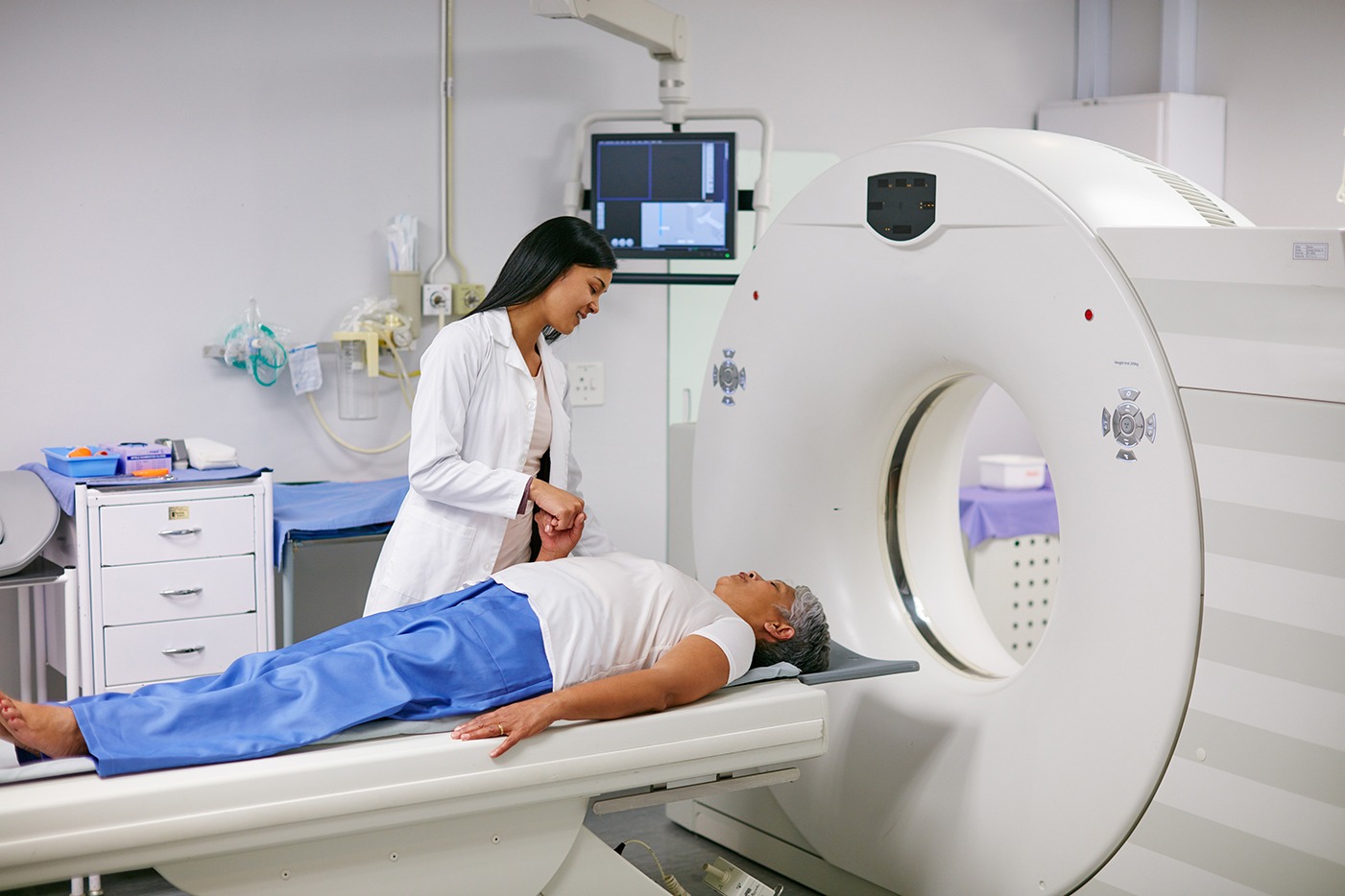
3] The MRI Scan: The patient will lie on an MRI table that is then moved into the MRI machine, a large cylindrical magnet. The MRI machine uses powerful magnetic fields and radio waves to create detailed images of the blood vessels. During the scan, the patient may be asked to hold their breath or stay still for short periods to avoid blurring the images. The scan usually lasts about 30 to 60 minutes, depending on the area being imaged.
4] Post-Procedure: After the MRI scan, the patient can resume normal activities immediately, unless they were given sedation during the procedure. The contrast dye is generally eliminated from the body through the kidneys and urine.


