Why is CT Aortography Important?
CT Aortography is a powerful diagnostic tool with the following key applications:
1. Assessment of Aneurysms: One of the most common reasons for performing CT aortography is to detect aortic aneurysms. An aneurysm is a bulge or enlargement in the aorta that can weaken the vessel wall. If an aneurysm ruptures, it can be life-threatening. A CT scan can identify the size, shape, and location of an aneurysm and help doctors determine the best treatment plan.
2. Detection of Aortic Dissection: An aortic dissection occurs when the inner layer of the aortic wall tears, leading to blood flowing between the layers of the vessel wall. This is a life-threatening emergency that requires prompt diagnosis and intervention. CT aortography can quickly visualize the tear and assess its severity.
3. Evaluation of Aortic Stenosis and Other Blockages: Aortic stenosis is a condition in which the aortic valve narrows, obstructing blood flow. CT aortography can help assess the degree of narrowing, as well as identify any blockages or narrowing in the aorta and its branches, such as those affecting the renal arteries or carotid arteries.
4. Preoperative Planning: In cases where surgery or interventional procedures, such as stent placement, are planned to treat aortic conditions, CT aortography provides detailed information about the aorta's anatomy. This information helps surgeons plan the best approach for the procedure.
5. Vascular Malformations: In addition to aneurysms and dissections, CT aortography is also useful in detecting vascular malformations or congenital abnormalities in the aorta and its branches. These conditions may require medical management or surgical correction.
6. Follow-up of Aortic Surgery: For patients who have undergone surgery or intervention on the aorta, CT aortography is often used for follow-up to ensure that the repaired area is healing well and that there are no new complications, such as the recurrence of an aneurysm or the development of other issues in the aorta.
For precise CT Aortography in Pune, Diagnopein offers advanced imaging services with cutting-edge technology. If you're searching for a CT scan centre near me, Diagnopein ensures accurate results and quick diagnostics. This test helps assess the aorta for conditions like aneurysms or blockages, offering essential insights for cardiovascular health. With expert care and reliable reports, Diagnopein is your trusted partner for comprehensive vascular assessments in Pune.
How is CT Aortography Performed?
The process for CT aortography involves several steps to ensure accurate imaging:
1. Preparation: Before the scan, the patient may need to remove clothing and any metallic items (such as jewelry or piercings) that could interfere with the imaging. Depending on the hospital's protocols, the patient may need to lie on a CT scanner bed and may be asked to fast for a few hours before the procedure if contrast dye is used.
2. Injection of Contrast Dye: The contrast dye, typically an iodine-based solution, is injected into a vein (usually in the arm) to enhance the visibility of the blood vessels. The contrast material makes the aorta and blood vessels appear brighter on the CT scan, allowing for better visualization of any abnormalities.
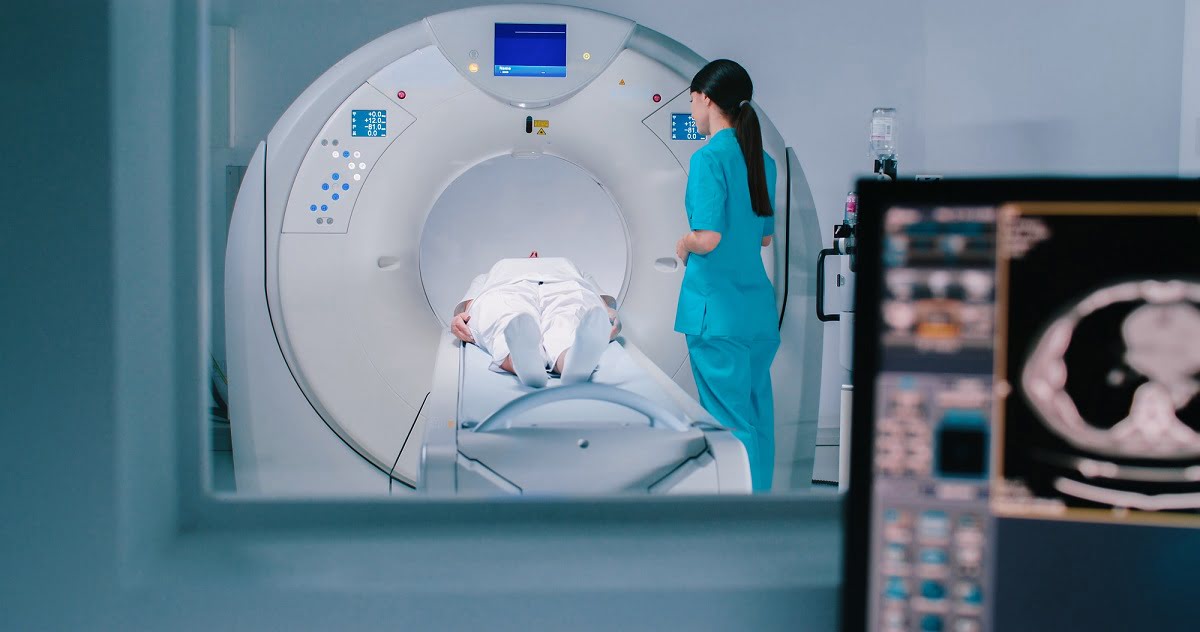
3. Imaging Process: Once the contrast dye is administered, the patient will be positioned on the CT scanner bed, which may move through the scanner’s large circular opening. The scanner will rotate around the patient, capturing multiple cross-sectional images of the aorta and surrounding structures. The images are processed by a computer to create detailed, 3D images of the aorta.
4. Breathing and Positioning: The patient may be asked to hold their breath for short periods during the scan to ensure the images are clear and free from movement. This is particularly important as movement can affect the quality of the images.
5. Post-Scan Care: After the procedure, the patient is typically monitored for a short time to ensure there are no reactions to the contrast dye. Patients who received contrast material may be asked to drink fluids to help flush the dye from their system. There are usually no other restrictions after the procedure, and patients can return to their normal activities.
Who Should Consider CT Aortography?
CT aortography is generally recommended for individuals who are at risk for or have symptoms of aortic diseases. These include:
1. Suspected Aortic Aneurysm or Dissection: Patients who experience severe chest, back, or abdominal pain, particularly if it is sudden or sharp, may be evaluated with CT aortography. It is commonly used in emergency situations when aortic aneurysm or dissection is suspected.
2. Vascular Disease: Individuals with a history of atherosclerosis (hardening of the arteries) or other vascular diseases may require CT aortography to assess the health of their aorta and related blood vessels. Those with high blood pressure, high cholesterol, or a family history of aortic conditions may also be candidates for this test.
3. Follow-up for Aortic Surgery: For patients who have previously had surgery or intervention for aortic conditions, CT aortography is used for routine follow-up to monitor for potential complications or new issues.
4. Preoperative Evaluation: CT aortography may be ordered prior to surgery or interventional procedures to assess the anatomy of the aorta and ensure that the surgical team has accurate information for planning the procedure.
5. Chest or Abdominal Pain: For patients with unexplained chest or abdominal pain, especially if the pain is severe, sudden, or associated with risk factors for cardiovascular diseases, CT aortography can help rule out or confirm aortic problems.


