What is MRI Venography? – Detailed Overview
MRI Venography (MRV) is a specialized magnetic resonance imaging procedure that produces high-resolution images of veins in various parts of the body, such as the brain, abdomen, pelvis, or legs. Unlike traditional venograms, MRV does not use radiation, making it safer and more patient-friendly.
This test is particularly important in diagnosing conditions like:
-
Deep vein thrombosis (DVT)
-
Cerebral venous thrombosis
-
Venous obstruction or narrowing
-
Varicose veins
-
Congenital vascular abnormalities
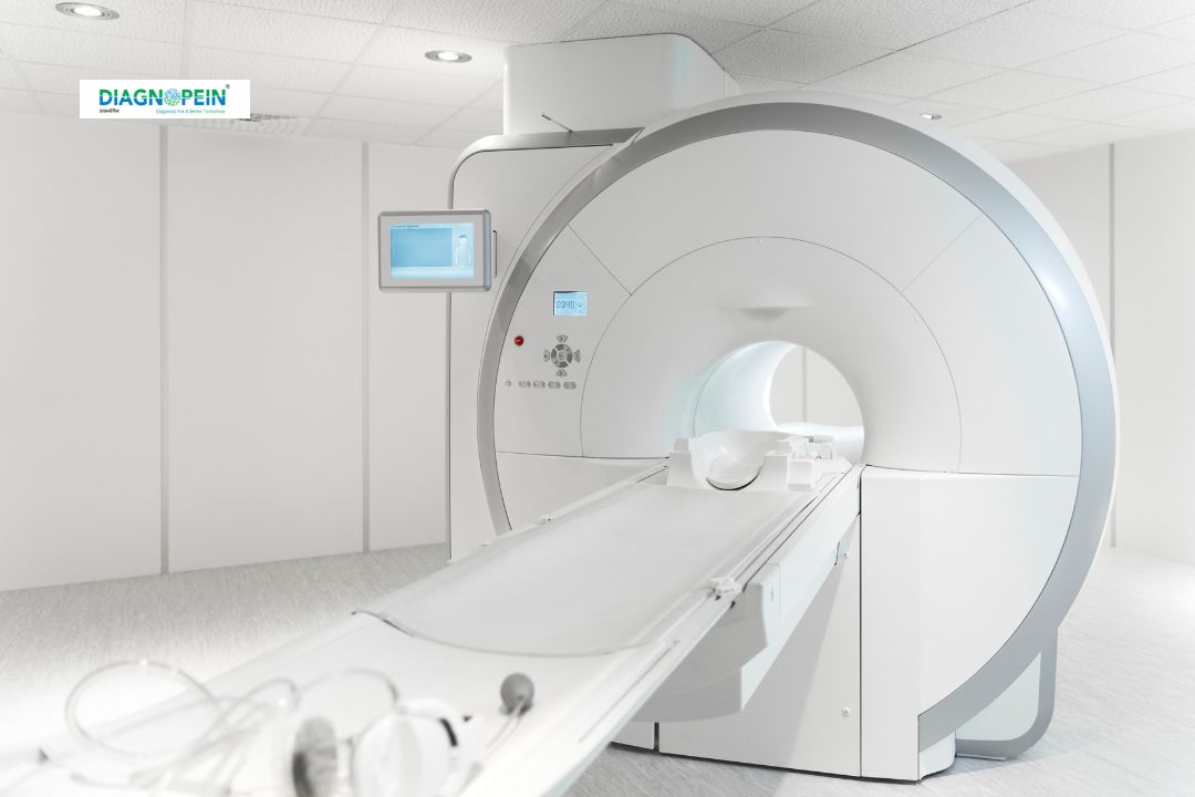
At Diagnopein, MRI Venography in Nanded is performed using advanced software and 3 Tesla MRI machines to ensure exceptional clarity, faster scans and improved diagnostic accuracy.
Why MRI Venography is Important?
Understanding venous health is crucial because veins carry deoxygenated blood back to the heart. Any blockage or clot can interrupt this flow and cause severe complications. MRI Venography plays an essential role in early detection and management of venous disorders.
Importance of MRI Venography:
-
Helps identify blood clots before they become life-threatening
-
Detects vein narrowing or blockages affecting blood circulation
-
Useful in diagnosing brain venous sinus thrombosis
-
Helps vascular surgeons plan treatment or surgery
-
Provides detailed imagery without radiation exposure
-
Helps track progress of existing venous conditions
Diagnosing these issues early through MRI Venography prevents complications like stroke, pulmonary embolism, and chronic vein insufficiency.
Benefits of MRI Venography
There are several reasons why doctors prefer MRI Venography over other vascular imaging techniques:
? Non-invasive and radiation-free
MRV uses magnetic fields and is safe for repeated evaluations.
? High-resolution 3D venous imaging
It generates detailed images from multiple angles for precise diagnosis.
? Detects both acute and chronic vein issues
MRV can identify new clots as well as old thrombotic formations.
? Safe for most patients
Suitable for children, pregnant women (when recommended), and elderly patients.
? Quick and highly accurate
Most scans are completed in 20–30 minutes with highly reliable results.
How the MRI Venography Test is Done? – Step-by-Step Procedure
At Diagnopein, we ensure a smooth, comfortable, and hygienic testing experience for every patient. The MRI Venography procedure includes:
Step 1: Pre-scan preparation
Patients are asked to remove metal objects. No major fasting is required unless specified.
Step 2: Positioning on the MRI table
The technologist positions the patient depending on the area being scanned.
Step 3: Use of contrast (when needed)
Some MRV studies require contrast to highlight veins more clearly. It is safe and routinely used.
Step 4: Image acquisition
The MRI machine captures detailed venous images using magnetic pulses.
Step 5: Interpretation by radiologist
Highly trained radiologists evaluate the images and prepare an accurate report.
MRI Venography Parameters
Typical parameters evaluated during MRI Venography include:
-
Venous flow direction and velocity
-
Vein patency (open or blocked)
-
Presence of thrombosis or clots
-
Vein size, shape, and structure
-
Venous sinus visualization (in brain MRV)
-
Collateral circulation assessment
-
Inflammatory changes in vein walls
These parameters help doctors understand the exact condition of the venous system and recommend the right treatment.
Why Choose Diagnopein for MRI Venography in Nanded?
Diagnopein offers advanced MRI scanning facilities with expert radiologists and accurate reporting. With high-end MRI systems, comfortable scanning rooms, and affordable pricing, we ensure reliable diagnosis for better treatment outcomes. Patients in Nanded prefer Diagnopein due to precision, transparency and world-class technology.