Overview of MRI Temporomandibular – B/L – Without Contrast
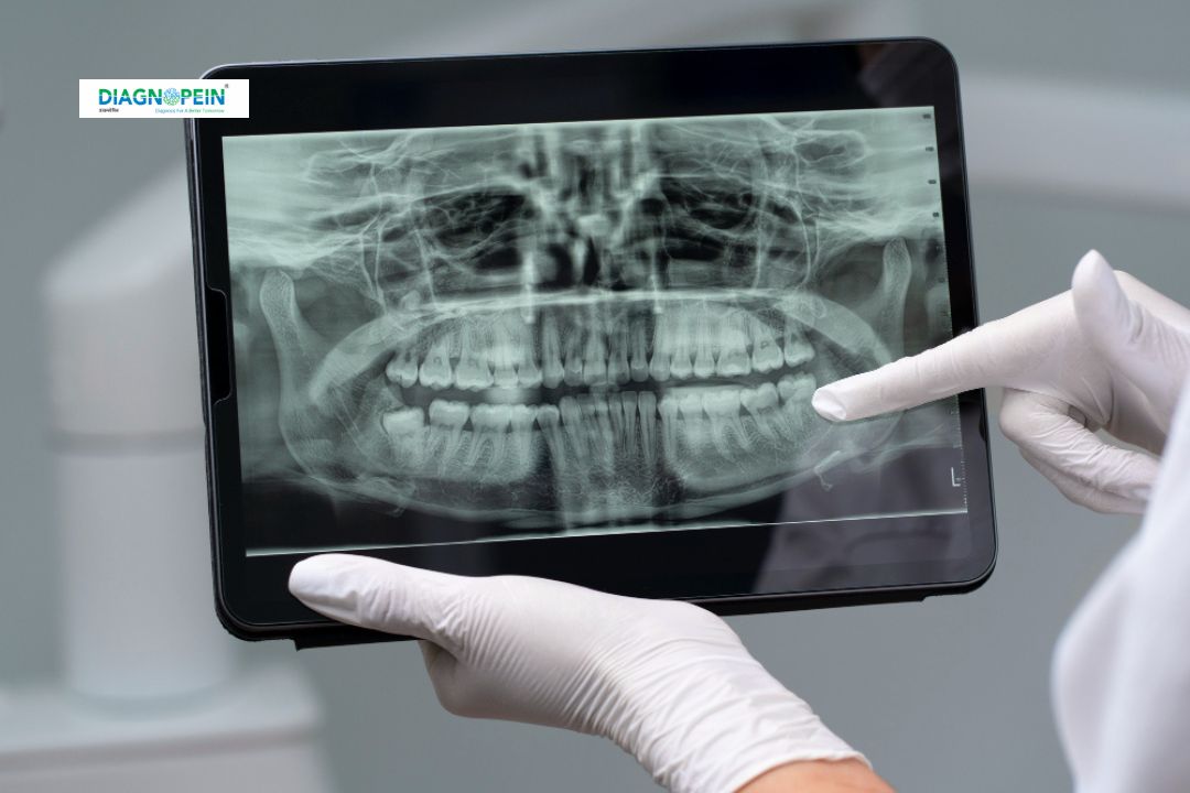
The MRI Temporomandibular – B/L – Without Contrast scan is performed to evaluate both TMJs simultaneously, ensuring a complete assessment of jaw structure and function. MRI is the gold standard for TMJ imaging because it shows both bone and soft-tissue details, including cartilage, disc position, joint effusion, and ligament integrity.
This technique is non-invasive, radiation-free, and extremely precise, making it ideal for diagnosing early-stage TMJ disorders before they worsen. At Diagnopein Nanded, this scan is performed using high-end MRI equipment ensuring instant and reliable reporting.
Why MRI Temporomandibular – B/L – Without Contrast Is Important
This MRI scan is essential for identifying the root cause of TMJ-related symptoms. Patients often present with jaw clicking, popping, locking, headaches, ear discomfort, and difficulty chewing. Since TMJ disorders involve soft tissues and disc displacement, MRI is the only imaging test capable of showing the internal structure clearly.
This test helps detect:
-
Disc displacement with or without reduction
-
Internal derangement of the TMJ
-
Joint inflammation or effusion
-
Degenerative joint disease (arthritis)
-
Trauma-related damage
-
Congenital or developmental abnormalities
Early diagnosis helps prevent complications and ensures better treatment outcomes. People searching for TMJ imaging or overview in Nanded can rely on Diagnopein for expert evaluation.
Benefits of MRI Temporomandibular – B/L – Without Contrast
Choosing MRI Temporomandibular – B/L – Without Contrast provides multiple benefits, including:
? Non-invasive and Radiation-free
Unlike CT scans or X-rays, MRI uses magnetic fields instead of radiation.
? Clear Visualization of Soft Tissues
Ideal for viewing disc position, ligaments, synovial fluid, and joint spaces.
? Bilateral Assessment in One Scan
Both joints can be evaluated simultaneously for symmetrical comparison.
? No Contrast Dye Required
Safe for all patients, including those with dye allergies or kidney concerns.
? Accurate Diagnosis for TMJ Treatment Planning
Helps dentists, orthodontists, ENT specialists, and maxillofacial surgeons create accurate treatment plans.
At Diagnopein Nanded, patients benefit from expert radiology reporting, advanced MRI systems, and comfortable scanning experience.
How the MRI Temporomandibular – B/L – Without Contrast Test Is Done
The procedure is simple, painless, and usually completed in 20–30 minutes.
Step-by-Step Process
-
The patient lies on the MRI table comfortably.
-
A coil is placed near the jaw area to capture clear TMJ images.
-
Multiple sequences are taken with the mouth closed and open positions.
-
No injections, dyes, or contrast are used.
-
Images are reviewed by expert radiologists for detailed reporting.
Patient Preparation
-
Remove jewelry, metallic objects, and dentures.
-
Inform the team if you have implants, pacemakers, or metal fragments.
-
No fasting or special preparation is required.
This makes the test safe, quick, and suitable for all age groups.
Parameters Covered in MRI Temporomandibular – B/L – Without Contrast
The scan typically includes:
- Proton density sequences
-
Open-mouth and closed-mouth imaging
-
Dynamic motion evaluation
-
Disc position analysis
-
Joint space measurement
-
Effusion detection
-
Soft-tissue and cartilage assessment
These parameters help radiologists deliver highly accurate findings for TMJ disorders.