Importance of X-Ray Wrist AP/LAT with Scaphoid View
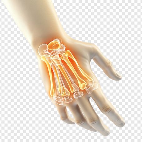
The wrist consists of multiple small bones that work together to allow smooth hand and wrist movement. The scaphoid bone connects both rows of carpal bones and is critical for wrist stability. Any injury or fracture, especially a missed scaphoid fracture, can cause long-term complications if not detected early.
The X-Ray Wrist AP/LAT with Scaphoid View is crucial because:
-
It identifies hidden scaphoid fractures that might not appear on standard wrist X-rays.
-
It helps detect subtle dislocations or bone misalignments.
-
It assists doctors in planning surgical or non-surgical treatments.
-
It ensures proper monitoring of healing after wrist surgery or casting.
-
It supports accurate orthopedic and physical therapy assessments.
For individuals experiencing chronic wrist pain or post-injury swelling, an X-Ray Wrist AP/LAT and Scaphoid view is an essential diagnostic step.
Benefits of X-Ray Wrist AP/LAT with Scaphoid View at Diagnopein
Choosing Diagnopein for your X-Ray Wrist AP/LAT with Scaphoid View ensures accuracy, comfort, and reliability. Our expert radiology team uses digital imaging to deliver detailed diagnostic reports with high clarity.
Key benefits include:
-
Fast and non-invasive imaging process
-
High-definition visuals of wrist and scaphoid bones
-
Early fracture detection to prevent complications
-
Minimal radiation exposure ensuring patient safety
-
Same-day results interpreted by expert radiologists
Our advanced imaging systems optimize diagnostic efficiency, enabling timely treatment and faster recovery for patients.
Procedure and Parameters of the Test
During the X-Ray Wrist AP/LAT with Scaphoid View, the patient is positioned carefully to obtain clear images of all essential wrist bones. Generally, three images are taken: anteroposterior (AP), lateral (LAT), and oblique or scaphoid view.
Procedure steps:
-
The technician positions the patient’s wrist on the X-ray table.
-
The hand may be slightly rotated or adjusted to focus on the scaphoid bone.
-
The radiographer captures multiple angles to ensure complete bone coverage.
-
The images are processed digitally and reviewed by a radiologist for reporting.
Common parameters considered:
-
Bone density and alignment
-
Joint spacing between carpal bones
-
Visible fractures or hairline cracks
-
Presence of dislocation or bone trauma
-
Clarity of the scaphoid and surrounding bones
Patients are advised to remove any metallic jewelry before the scan to prevent image distortion. The test usually takes less than 10 minutes and requires no special preparation.