Importance of X-Ray Townes AP View
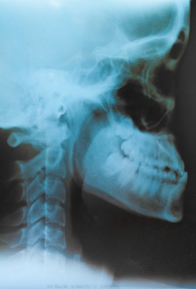
The Townes AP X-Ray plays a crucial role in neuro and orthopedic diagnostics. It provides a clear view of the occipital bone and posterior fossa, which are difficult to assess in standard anteroposterior (AP) or lateral skull views. Doctors recommend this test when they suspect:
-
Skull base or occipital bone fractures
-
Congenital deformities of the craniovertebral junction
-
Tumors or bone lesions near the foramen magnum
-
Post-surgical follow-up after cranial or cervical interventions
By offering a precise image of the posterior skull base, the Townes view aids surgeons and neurologists in accurate diagnosis and treatment planning.
Benefits of X-Ray Townes AP View
An X-Ray Townes View test at Diagnopein offers multiple benefits that make it a preferred choice for clinicians and patients alike:
-
High clarity of skull base structures: Enhanced viewing of the occipital bone, mastoid, and foramen magnum.
-
Quick and non-invasive procedure: The entire process takes less than 10 minutes with no discomfort to the patient.
-
Accurate diagnostic aid: Enables early detection of fractures, sinus pathology, and cranial deformities.
-
Affordable and accessible: Diagnopein provides cost-effective skull X-ray services accessible to residents of Karad and surrounding areas.
-
Minimal radiation exposure: Digital X-ray systems reduce exposure while ensuring precise images.
How X-Ray Townes AP View Test is Performed
The Townes view X-Ray procedure is performed under expert supervision using a standard protocol to ensure accuracy and patient safety.
-
The patient is seated upright or lying supine.
-
The head is positioned so that the orbitomeatal line is at an angle of about 30° to the X-ray beam.
-
The beam passes through the occiput toward the foramen magnum at an anterior-posterior (AP) angle.
-
The radiographer ensures no movement during exposure to prevent blurring.
-
Digital images are then evaluated by radiologists for diagnostic interpretation.
This specialized setup helps produce a clear projection of the occipital bone and upper cervical region, revealing minute details.
Parameters and Technical Factors
To achieve optimum image quality, specific exposure parameters are maintained during the Townes AP X-Ray:
-
Position: AP projection with head flexed about 30° from the horizontal plane
-
Central ray: Directed through the foramen magnum
-
Film focus distance: Approximately 100 cm
-
kVp range: 70–80 kVp depending on patient build
-
mAs settings: Optimized for high contrast and low noise
-
Collimation: Restricted to the area of interest to reduce scatter and patient dose
These parameters ensure diagnostic accuracy and safety compliance following radiographic standards.
Why Choose Diagnopein for X-Ray Townes AP View
Diagnopein stands out for its commitment to quality imaging and patient care. With modern digital X-ray systems, trained radiologic technologists, and prompt reporting, Diagnopein has become a trusted diagnostic center in Karad.
Patients and physicians rely on our imaging reports for precise evaluation of head and skeletal conditions. Efficient service, hygienic environment, and expert assistance make Diagnopein your dependable partner for X-Ray Townes AP View and other radiological tests.