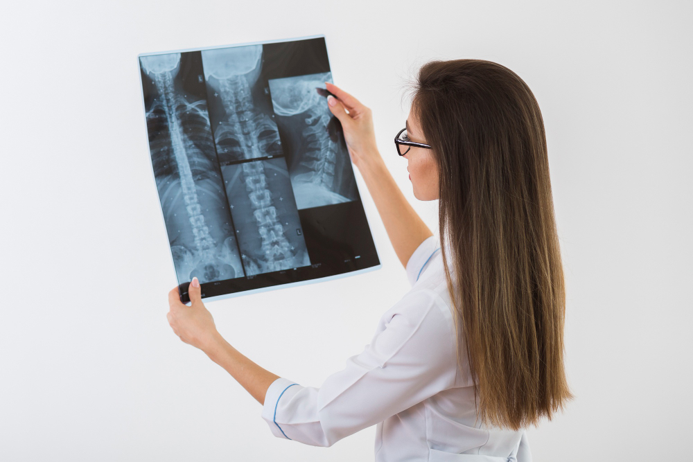
Importance of X-Ray TM Joint Lateral View
The X-Ray TM Joint Lateral View is important for identifying structural or functional abnormalities in the jaw joints. It provides detailed insight into the relationship between the mandibular condyle and the temporal bone, especially during opening and closing of the mouth.
This test helps in diagnosing:
-
Temporomandibular joint disorders (TMD)
-
Dislocation or subluxation of the joint
-
Degenerative joint diseases like arthritis
-
Fractures involving the TMJ area
-
Post-surgical evaluation or trauma follow-up
Accurate TMJ imaging ensures early treatment and helps prevent chronic pain, headaches, and jaw stiffness associated with untreated conditions.
Benefits of X-Ray TM Joint Lateral View
Undergoing a TM Joint Lateral X-Ray offers several clinical and diagnostic advantages:
-
Helps detect subtle joint abnormalities that may not be visible externally
-
Non-invasive, quick, and painless procedure
-
Enables accurate planning for dental surgeries or orthodontic corrections
-
Assists in monitoring post-operative healing of TMJ-related procedures
-
Useful for evaluating jaw alignment and mouth-opening capacity
These benefits make the X-Ray TM Joint Lateral View a crucial part of comprehensive TMJ evaluation and treatment planning, especially for patients experiencing persistent jaw discomfort or clicking sounds while opening the mouth.
4. How X-Ray TM Joint Lateral View Test is Done
The TM Joint X-Ray Lateral View procedure at Diagnopein is performed by experienced radiology professionals following all standard safety protocols.
Here’s how the test is done:
-
The patient is positioned carefully with one side of the face against the X-ray plate.
-
The technologist ensures the jaw is in a closed, open, or partially open position, depending on the prescribed evaluation.
-
The X-ray machine emits a controlled beam toward the TM Joint area, capturing detailed side-view images.
-
The images are then processed and evaluated by radiologists for bone movement, spacing, and joint shape analysis.
The entire process takes only a few minutes and uses minimal radiation exposure to ensure patient safety.
Parameters Assessed in TMJ Lateral View
The X-Ray TM Joint Lateral View helps assess several diagnostic parameters critical to evaluating joint health:
-
Condylar position and shape
-
Joint space width
-
Glenoid fossa structure
-
Range of mandibular motion
-
Presence of osteoarthritic changes
-
Bilateral symmetry of TMJ functions
Radiologists analyze these readings to differentiate between normal and pathological conditions, guiding dentists or maxillofacial surgeons toward accurate treatment decisions.
Why Choose Diagnopein for TMJ X-Ray in Karad
Diagnopein offers high-quality TM Joint X-Ray services in Karad, combining advanced imaging technology with expert radiologists. We ensure clear, reliable results in minimal time with maximum patient comfort. Our focus on precision and care makes us a trusted choice for dental and maxillofacial imaging.
Whether you are experiencing jaw stiffness, facial pain, or clicking sounds in your joint, Diagnopein’s TMJ X-Ray test helps uncover the cause quickly and accurately.