


Diagnopein X-RAY THORACIC SPINE LATERAL VIEW Centre in Karad
Diagnopein X-RAY THORACIC SPINE LATERAL VIEW Centre in Karad
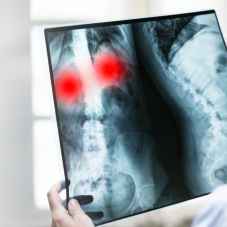
The X-Ray Thoracic Spine Lateral View is a diagnostic imaging test that captures detailed side-view images of the thoracic (mid-back) portion of the spine. This test helps visualize the vertebrae, intervertebral spaces, and surrounding soft tissues to detect bone deformities, spinal alignment issues, or disc-related problems.
At Diagnopein Diagnostic Center, Karad, modern digital X-ray technology ensures high-quality imaging with minimal radiation exposure, making it safe, quick, and comfortable for patients.
Common related searches include thoracic spine lateral X-ray, X-ray of middle back, and spine side view X-ray — all referring to the same imaging procedure performed with precision at Diagnopein.


