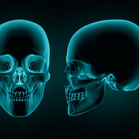
Importance of X-Ray Skull Lateral
The X-Ray Skull Lateral view plays a crucial role in evaluating structural damage and abnormalities of the skull. It helps radiologists and neurologists identify fractures, brain swelling, and sinus blockages that may not be visible during physical examinations.
Key importance includes:
-
Detecting skull fractures, injuries, and bone deformities.
-
Evaluating sinus cavities for infections or inflammation.
-
Assessing the position of surgical implants or medical devices.
-
Supporting the diagnosis of congenital skull anomalies.
-
Acting as a preliminary investigation before advanced scans like CT or MRI.
At Diagnopein, the X-Ray Skull Lateral scan provides precise diagnostic insights that assist in faster and more accurate medical decision-making.
Benefits of X-Ray Skull Lateral
Undergoing a Skull Lateral X-Ray offers several patient advantages, especially for emergency care and detailed bone assessment. Some of the main benefits include:
-
Quick and simple process: The test takes only a few minutes and requires minimal preparation.
-
Non-invasive and painless: No incisions or discomfort; it uses safe, low-dose radiation.
-
Supports accurate treatment: Helps your doctor plan surgical or medical treatment effectively.
-
Affordable and widely available: Compared to MRI or CT scans, lateral X-rays are cost-effective and easy to access.
-
High diagnostic value: Useful in detecting even minor fractures, sinus infections, or brain-related concerns.
At Diagnopein Karad, we ensure radiation safety, patient comfort, and accurate results through state-of-the-art X-Ray technology.
How X-Ray Skull Lateral Test is Done
The X-Ray Skull Lateral test procedure is performed by trained radiographers following strict safety guidelines. The process includes:
-
The patient is asked to remove metal objects such as earrings or glasses.
-
The radiographer positions the patient’s head sideways (lateral view).
-
The X-ray machine focuses on the skull while maintaining the correct distance and angle.
-
The patient remains still for a few seconds as the image is captured.
-
The radiologist examines and interprets the digital image for diagnosis.
The entire test usually completes within 10–15 minutes, and results are provided digitally or in print within a short time frame.
Parameters and Technical Insights
During a Skull Lateral X-Ray, several parameters and technical factors ensure clear image quality and diagnostic reliability. These parameters include:
-
Field of view (FOV): Covers the entire skull.
-
Exposure factors: Adjusted based on patient age and anatomy.
-
Image contrast: Optimized to evaluate bone and soft tissue structures.
-
Radiation dose: Kept to the minimum, complying with safety standards.
-
Positioning accuracy: Essential for distinguishing cranial features.
At Diagnopein, we use modern radiography systems that enhance image clarity and reduce noise, ensuring reliable results for all patients.
Why Choose Diagnopein for X-Ray Skull Lateral in Karad
Choosing Diagnopein means choosing accuracy, comfort, and care. Our diagnostic center in Karad is equipped with digital X-Ray technology, experienced radiologists, and safe imaging environments. Whether it’s a skull injury evaluation or routine check, we provide dependable imaging services backed by efficient reporting and patient support.
We prioritize patient safety, precise diagnosis, and quick result delivery—making Diagnopein the trusted destination for X-Ray Skull Lateral in Karad.


