Why X-Ray Skull AP/Lateral is Important
X-Ray Skull AP/Lateral plays a crucial role in diagnosing and monitoring several medical conditions related to the head and cranial region. It allows doctors to visualize internal bone structures without the need for invasive procedures. The test is particularly important to:
-
Identify skull fractures after head injury
-
Detect sinus infections or fluid accumulation
-
Evaluate congenital skull deformities and bone lesions
-
Monitor post-surgical bone healing
-
Detect foreign objects or calcifications in the head region
At Diagnopein, the Skull X-Ray AP/Lateral test helps clinicians get early and accurate insights into cranial abnormalities, supporting effective treatment planning and patient recovery.
Benefits of X-Ray Skull AP/Lateral
Getting a Skull AP/Lateral X-Ray offers several diagnostic and safety benefits. It provides high-quality images in minutes, allowing swift medical evaluation and treatment options.
Main Benefits Include:
-
Detailed Cranial Assessment: Two-view imaging helps radiologists evaluate skull alignment, bone injuries, and tissue conditions accurately.
-
Quick and Non-Invasive: No needles or anesthesia required—patients experience a short, comfortable imaging process.
-
Minimal Radiation Exposure: Using advanced digital radiography, Diagnopein minimizes exposure for all age groups.
-
Early Disease Detection: Allows doctors to detect infections, abnormal growths, or injuries before they worsen.
-
Accurate Post-Injury Analysis: Useful for follow-ups after head trauma or surgery.
These benefits make X-Ray Skull AP/Lateral a safe and effective diagnostic option for both adults and children.
Procedure and Parameters of X-Ray Skull AP/Lateral
The procedure for an X-Ray Skull AP/Lateral at Diagnopein is quick, simple, and handled by experienced radiology technicians. The patient is positioned correctly to ensure high-quality images with precision.
Procedure Steps:
-
The patient removes any metal objects or jewelry from the head and neck area.
-
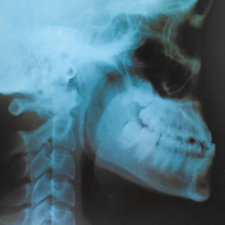
For the AP (Anteroposterior) view, the patient’s face is positioned facing the X-ray beam directly.
-
For the Lateral view, the patient’s head is turned sideways to capture a detailed side image.
-
The technician adjusts exposure parameters like kilovoltage (kV), milliamperes (mA), and exposure time based on the patient’s age and build.
-
The scan takes only a few minutes, and the results are processed digitally for quick reporting.
Key Parameters:
-
Views: AP & Lateral
-
Exposure: Adjusted per patient
-
Duration: 5–10 minutes
-
Radiation Dose: Low, safe level
-
Result Time: Within 15–30 minutes
At Diagnopein Karad, patients receive accurate Skull X-Ray AP/Lateral reports through advanced imaging and expert radiologist analysis, ensuring timely diagnosis and treatment.