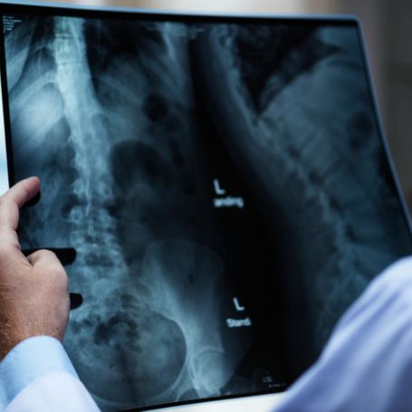
Why X-Ray SI Joint Is Important
The sacroiliac joint is often overlooked when diagnosing back pain, but it plays a major role in body movement and posture. Pain or stiffness in this region could indicate several conditions such as sacroiliitis, ankylosing spondylitis, osteoarthritis, or trauma.
An X-Ray SI Joint test helps doctors identify the exact cause of pain or discomfort by revealing joint space narrowing, bone changes, or inflammation. Early detection through X-ray imaging enables proper medical intervention and prevents further complications.
Key diagnostic uses include:
-
Evaluating lower back and pelvic pain origins
-
Detecting joint inflammation and degeneration
-
Identifying arthritis or spondyloarthropathy
-
Monitoring post-injury healing or trauma
By performing this test at Diagnopein, patients receive accurate imaging that supports clinical decision-making and long-term treatment planning.
Benefits of Doing X-Ray SI Joint
At Diagnopein Diagnostic Center, we ensure every patient gets precise and safe imaging with quick report turnaround. The main benefits of an SI joint X-ray include:
-
Accurate diagnosis: Detects structural changes or bone erosion early.
-
Non-invasive test: Requires no injections or contrast mediums.
-
Quick results: X-ray imaging typically takes less than 10 minutes.
-
Clinical guidance: Assists doctors in determining treatment paths for pain management or physiotherapy.
-
Cost-effective: One of the most affordable diagnostic imaging options for joint issues.
This test is especially beneficial for individuals suffering from chronic back stiffness, unexplained hip pain, or suspected sacroiliitis.
How X-Ray SI Joint Test Is Done
The X-Ray SI joint procedure is simple, comfortable, and completed in minutes. Here’s how it is performed at Diagnopein:
-
The patient is positioned either supine (lying on the back) or slightly rotated to capture the best angle of both joints.
-
The X-ray beam is directed toward the sacroiliac region to get a clear view of joint spaces.
-
Our radiographer ensures accurate alignment, clarity, and appropriate exposure.
-
The generated digital images are reviewed and interpreted by experienced radiologists to identify any abnormalities or inflammation.
Patients do not need fasting or special preparation before the test. However, those with metallic implants should inform the radiologist beforehand.
Parameters and Report Interpretation
An SI joint X-ray report includes crucial parameters that help doctors evaluate joint health, such as:
-
Symmetry between right and left sacroiliac joints
-
Joint space width and alignment
-
Bone density and sclerosis signs
-
Early fusion or erosion patterns
-
Indications of arthritis or inflammation
At Diagnopein, our radiology experts analyze every detail with precision, ensuring you get accurate findings for further treatment.


