What is X-RAY RIGHT KNEE AP/LAT/OBLIQUE?
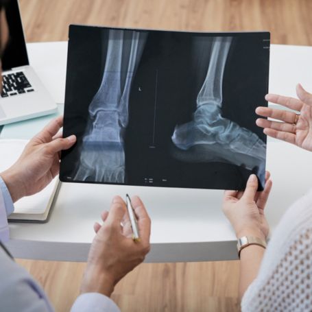
The X-RAY RIGHT KNEE AP/LAT/OBLIQUE is a radiographic examination of the right knee taken from three standard views—anteroposterior (AP), lateral (LAT), and oblique angles.
-
The AP view shows the front-to-back alignment of the knee joint.
-
The Lateral view focuses on the side profile of bones and surrounding structures.
-
The Oblique view provides a detailed angle between the front and side to capture hidden fractures or soft tissue changes.
This multi-angle approach allows doctors to get a 3D understanding of the knee’s internal condition, making diagnosis more accurate and complete.
Importance of X-RAY RIGHT KNEE AP/LAT/OBLIQUE
Performing an X-RAY RIGHT KNEE AP/LAT/OBLIQUE is essential in various medical conditions. It helps detect issues that a single-view X-ray might miss.
Major diagnostic uses:
-
Detecting fractures after accidents or falls
-
Evaluating joint alignment and bone deformities
-
Monitoring post-surgical or post-fracture healing
-
Identifying degenerative diseases like osteoarthritis
-
Locating foreign bodies or bone spurs
At Diagnopein Karad, every X-RAY RIGHT KNEE AP/LAT/OBLIQUE is performed under expert supervision, ensuring patient comfort and precise imaging.
Benefits of X-RAY RIGHT KNEE AP/LAT/OBLIQUE at Diagnopein Karad
Choosing Diagnopein Karad for your knee X-ray ensures high accuracy, faster reporting, and expert analysis. Here’s why patients trust us:
-
High-Resolution Digital Imaging: Crisp images that aid accurate diagnosis.
-
Quick Reporting: Same-day results to begin prompt treatment.
-
Minimal Radiation Exposure: Advanced low-dose technology for patient safety.
-
Comfortable Experience: Well-trained technicians and efficient service.
-
Comprehensive Assessment: The AP, Lateral, and Oblique views together allow doctors to identify even small fractures or early degenerative changes.
Our diagnostic expertise and cutting-edge equipment make Diagnopein Karad one of the most trusted centers for X-RAY RIGHT KNEE AP/LAT/OBLIQUE.
How is the X-RAY RIGHT KNEE AP/LAT/OBLIQUE Test Performed?
The process of performing an X-RAY RIGHT KNEE AP/LAT/OBLIQUE is simple and takes only a few minutes:
-
The patient is positioned on the X-ray table with the right knee placed appropriately for each view.
-
The technologist captures three images – AP (front view), LAT (side view), and Oblique (angled view).
-
Patients are asked to remain still to avoid image blur.
-
Once completed, the images are processed digitally and reviewed by radiologists.
No special preparation is required for the test. Patients are advised to remove any metal objects near the knee area before imaging.
Parameters and What the X-Ray Shows
The X-RAY RIGHT KNEE AP/LAT/OBLIQUE evaluates several key parameters:
-
Bone continuity and alignment
-
Joint space between femur, tibia, and patella
-
Signs of fracture, dislocation, or bone lesions
-
Presence of arthritis or osteophyte formation
-
Soft tissue swelling or fluid accumulation
These findings help orthopedic specialists determine the cause of pain, stiffness, or injury and plan the best treatment or surgery accordingly.
Why Choose Diagnopein Karad for X-RAY RIGHT KNEE AP/LAT/OBLIQUE
-
Trusted and certified diagnostic center in Karad
-
Skilled technicians and radiologists
-
Comfortable and hygienic testing environment
-
Quick appointment scheduling and convenient reporting options
At Diagnopein Karad, every X-RAY RIGHT KNEE AP/LAT/OBLIQUE combines advanced imaging technology with compassionate care for the most reliable diagnosis.