Importance of X-Ray Pelvis AP
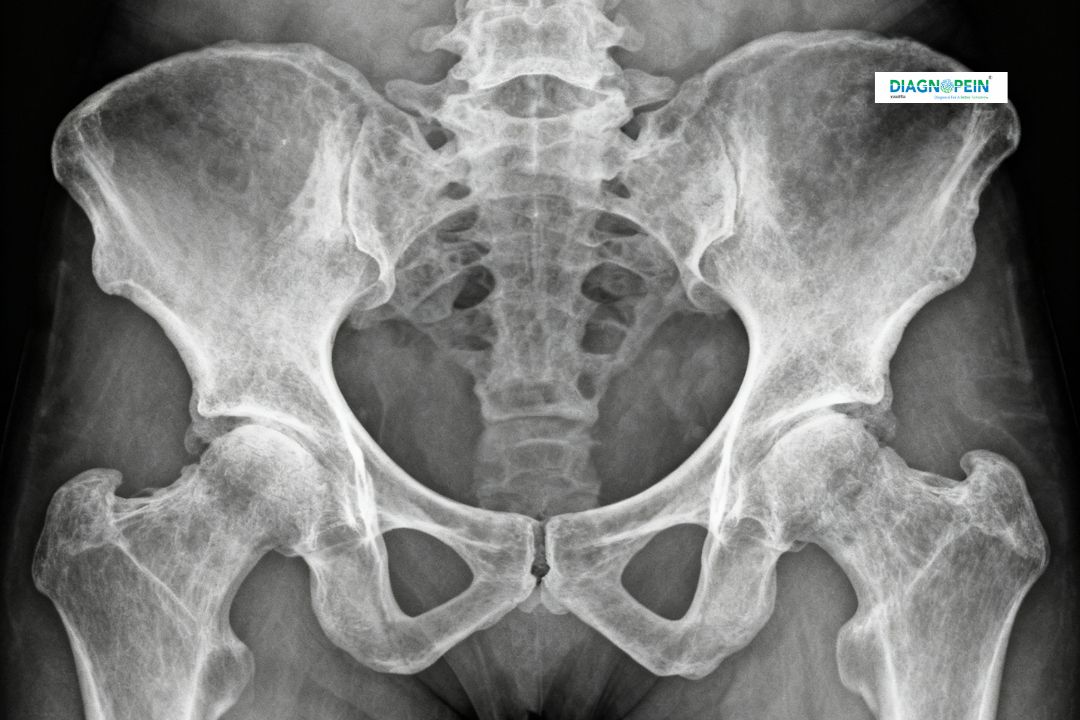
The X-Ray Pelvis AP is a critical diagnostic tool for evaluating injuries, joint degeneration, or bone disorders within the pelvic area. It helps detect fractures, dislocations, bone deformities, arthritis, or infections. For patients with lower back or hip pain, this scan helps physicians determine the root cause effectively.
At Diagnopein in Karad, doctors often recommend an X-Ray Pelvis AP for:
-
Detecting pelvic and hip fractures after an accident or fall
-
Evaluating hip dislocation or joint misalignment
-
Monitoring arthritis and degenerative joint conditions
-
Assessing healing progress after orthopedic surgery
-
Identifying any bone infections or tumors
With our high-quality imaging and expert reporting, patients in Karad receive accurate and timely results to support effective treatment decisions.
Benefits of X-Ray Pelvis AP
Getting an X-Ray Pelvis AP at Diagnopein Karad offers several benefits. The procedure is quick, painless, and non-invasive. It requires no special preparation and provides detailed images almost instantly. Some key benefits include:
-
Early detection of fractures and bone infections
-
Fast and safe imaging using low radiation exposure
-
Digital reporting for easy sharing and follow-up
-
Reliable tool for orthopedic, trauma, and post-surgical assessments
Our radiologists at Diagnopein in Karad ensure you get accurate diagnostics in minimal time, helping doctors plan the right course of treatment.
How the X-Ray Pelvis AP Test is Done
The procedure for an X-Ray Pelvis AP is simple and comfortable. The patient is asked to lie flat on the X-ray table with legs slightly rotated inward to get a proper front (anteroposterior) view of the pelvis. A lead apron is used to shield other parts of the body from unnecessary radiation exposure. Once positioned, the X-ray beam passes through the pelvis area while images are captured digitally. The entire process usually takes less than 10 minutes.
At Diagnopein Karad, the process is performed by trained radiologic technologists under strict safety standards. The reports are then interpreted by experienced radiologists to ensure precise diagnosis and quick report delivery.
Parameters and Technical Notes
-
Test Name: X-Ray Pelvis AP (Anteroposterior View)
-
Radiation Type: Low-dose digital X-ray
-
Duration: 5–10 minutes
-
Preparation: Usually no fasting or special preparation required
-
Safety: Safe for adults; special precautions applied for pregnant women
-
Report Time: Within a few hours
Patients can easily book their X-Ray Pelvis AP appointment at Diagnopein Karad online or by visiting our center directly. We prioritize patient convenience, prompt services, and diagnostic precision with every test conducted.