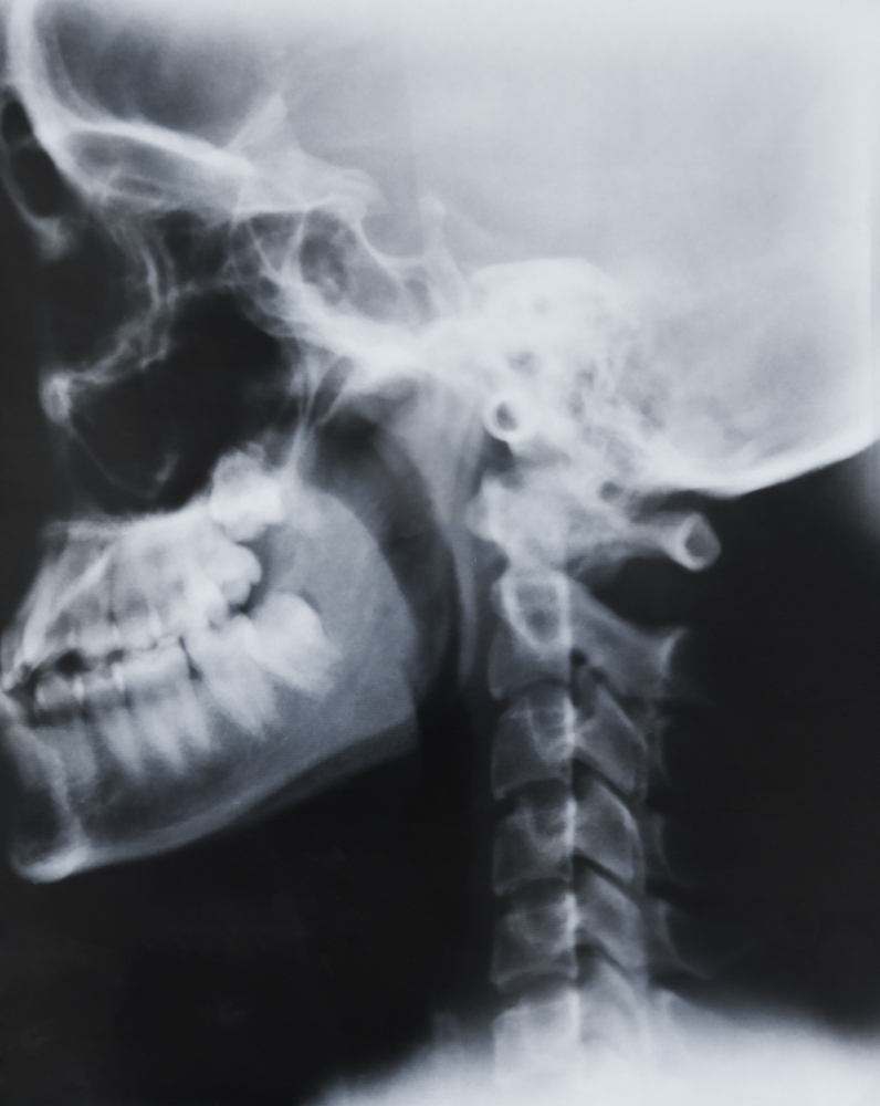
Why X-Ray Left Mastoid Schüller’s View is Important
This X-ray is crucial for evaluating chronic ear infections, mastoiditis, cholesteatoma, and post-surgical mastoid cavities. The Left Mastoid Schüller’s View allows radiologists to visualize the detailed anatomy of the temporal bone and its relationship to the middle and inner ear structures.
Key diagnostic purposes include:
-
Detecting inflammation or infection in the mastoid air cells.
-
Assessing bone destruction or sclerosis due to chronic otitis media.
-
Evaluating postoperative mastoid cavities after surgery.
-
Locating foreign bodies or abnormal growths behind the ear.
For patients in karad experiencing persistent ear pain, discharge, or hearing issues, this X-ray view can help in early diagnosis and prevent complications.
Benefits of X-Ray Left Mastoid Schüller’s View
The key advantages of this test include precision, speed, and affordability. At Diagnopein, our digital imaging technology ensures sharp and clear visuals, minimizing exposure time and radiation dose.
Major benefits:
-
Quick, non-invasive diagnostic process.
-
High-detail imaging of bony ear structures.
-
Useful for pre-surgical and post-operative mastoid evaluation.
-
Cost-effective compared to CT or MRI for initial screening.
Because mastoid investigations often require a specific angle, the Schüller’s View offers an optimal image projection, giving doctors an improved view of the left mastoid bone and helping them plan treatment accurately.
How the X-Ray Left Mastoid Schüller’s View Test is Performed
At Diagnopein in karad, the X-ray procedure is safe, simple, and comfortable for the patient. The technician positions the patient with the left side of the skull against the X-ray plate, tilting and angling the head slightly to achieve the ideal projection.
Steps include:
-
The patient removes metal objects and sits or stands near the X-ray unit.
-
The radiographer positions the head at a 25–30-degree angle to align the left mastoid properly.
-
The X-ray beam is directed toward the mastoid region with precision.
-
The image is captured digitally and reviewed immediately by a radiologist.
The entire process takes only a few minutes, and patients can resume normal activities right after. The results are provided promptly for consultation with the ENT specialist or surgeon.
Parameters and Diagnostic Focus
While performing the X-Ray Left Mastoid Schüller’s View, radiologists evaluate several parameters:
-
Clarity and aeration of mastoid air cells.
-
Integrity and thickness of the mastoid cortex.
-
Visibility of the external auditory canal and petrous ridge.
-
Bone density variations or erosions.
These details allow accurate analysis of any inflammatory or destructive changes, providing rich diagnostic data to support medical decision-making.
Why Choose Diagnopein in Karad
Diagnopein in karad stands out for its precision imaging technology, patient-centered care, and expert radiology staff. Our imaging center ensures minimal radiation exposure while maintaining image quality for detailed bone structure visualization.
We focus on providing fast, reliable reports that assist ENT and surgical specialists in planning effective treatments. Trusted by patients across karad, Diagnopein is committed to accurate diagnosis and compassionate care in every test we conduct.


