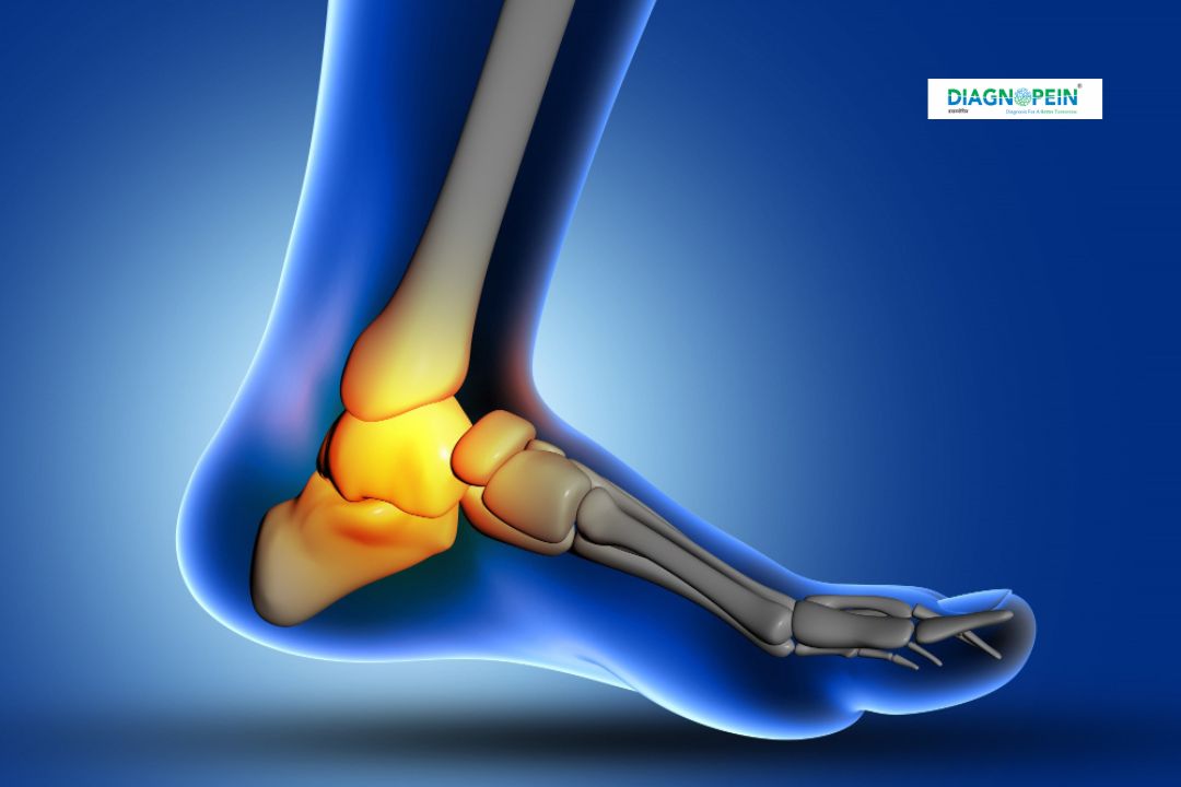
Why X-Ray Left Foot AP is Important
This test plays a crucial role in orthopedic and trauma diagnosis. When pain, bruising, or discomfort develops in the foot, an AP view X-ray helps determine whether there is a fracture, dislocation, or infection. It also assists in monitoring post-surgical outcomes and treatment progress.
In karad, Diagnopein offers accurate X-Ray Left Foot AP services backed by certified radiologists and experienced technicians. This diagnostic process supports the doctor’s ability to detect issues like bone tumors, gout, or osteoarthritis early, reducing the risk of future complications.
Key diagnostic insights from a Left Foot AP X-ray include:
-
Identifying bone fractures or cracks
-
Assessing joint spaces for signs of arthritis
-
Evaluating alignment abnormalities in the foot structure
-
Detecting infections, cysts, or bone lesions
Benefits of X-Ray Left Foot AP
An X-Ray Left Foot AP offers various health and diagnostic advantages. It is a painless, quick, and safe imaging procedure with minimal radiation exposure. Patients benefit from fast reporting, which allows for prompt medical intervention when necessary.
Key Benefits Include:
-
Non-invasive and risk-free diagnostic tool
-
High-resolution imaging for clear bone detail
-
Early detection of fractures or bone disease
-
Helpful in tracking healing post fracture or surgery
-
Immediate results for quicker treatment planning
At Diagnopein in karad, patients appreciate our fast, accurate, and affordable diagnostic imaging service. Whether for sports injuries or chronic foot pain, our advanced X-Ray Left Foot AP captures every anatomical detail to assist in precise diagnosis.
How X-Ray Left Foot AP Test is Done
The procedure for X-Ray Left Foot AP is simple and requires no special preparation. Patients may be asked to remove footwear and metal objects before the scan. The radiology technician positions the left foot on the X-ray table, ensuring the foot faces forward (AP view).
During the test:
-
The technician positions the foot correctly to capture the proper AP angle.
-
The X-ray beam passes from the front of the foot to the back.
-
The image is instantly captured using a digital detector or film.
-
Results are evaluated by a radiologist and shared with your doctor.
The entire process is completed within a few minutes, with minimal discomfort. At Diagnopein in karad, patient safety and image precision are our top priorities, ensuring that every test meets the highest medical standards.
Parameters and Diagnostic Value
The X-Ray Left Foot AP focuses on multiple diagnostic parameters, such as bone density, alignment, and joint spacing. It provides detailed evaluation of tarsal, metatarsal, and phalangeal bones. Radiologists assess these parameters to detect fractures, deformities, or degenerative changes.
Diagnostic Parameters Analyzed:
-
Bone integrity and alignment
-
Joint space width
-
Signs of inflammation or infection
-
Soft tissue density and calcification patterns
These parameters help doctors create a targeted treatment plan based on the underlying cause of pain, trauma, or deformity.
Choose Diagnopein in Karad for Trusted X-Ray Services
For reliable and precise X-Ray Left Foot AP imaging in karad, Diagnopein is your trusted diagnostic center. We combine advanced radiology technology, skilled technicians, and patient-centric care to deliver the best diagnostic outcomes. Our center also offers a complete range of imaging tests, from digital X-rays to ultrasound and MRI scans, ensuring comprehensive care under one roof.
Whether you’re dealing with post-injury assessment or routine bone health tracking, Diagnopein provides accurate reports that enable better medical decisions and faster recovery.