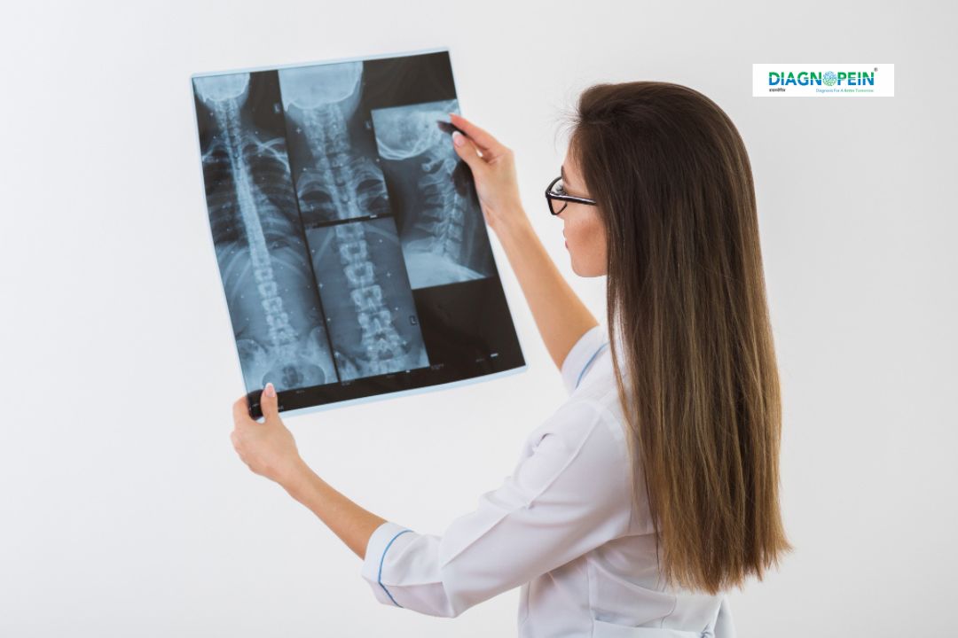
Why X-Ray Left Elbow Oblique View is Important
An X-Ray Left Elbow Oblique View is essential when a patient experiences elbow pain, swelling, restricted movement, or after trauma such as a fall or accident. The oblique angle captures overlapping bones and joint surfaces in detail, helping detect subtle fractures or alignment issues.
Common conditions detected through this imaging include:
-
Radial head fractures
-
Olecranon process injuries
-
Dislocations or bone fragmentation
-
Arthritis or joint degeneration
-
Post-operative assessment
At Diagnopein, karad, our radiology experts ensure every X-ray is performed under safe radiation guidelines, maintaining patient comfort and accuracy in each scan. The procedure is quick, non-invasive, and provides immediate visual evidence for further medical evaluation.
Benefits of Getting an X-Ray Left Elbow Oblique View
Choosing an X-Ray Left Elbow Oblique View at Diagnopein in karad comes with multiple benefits:
-
Clear visualization of complex elbow structures from an angled perspective
-
Early detection of hidden fractures and soft tissue abnormalities
-
Quick turnaround time with same-day report availability
-
Accurate diagnosis for effective treatment plans
-
Affordable pricing and easy appointment scheduling
Our diagnostic center in karad is equipped with latest-generation digital radiology machines that deliver minimal radiation exposure while producing sharp and detailed images. Trained radiologists interpret every scan with precision, ensuring reliable results for orthopedic surgeons, general practitioners, and physiotherapists.
How the X-Ray Left Elbow Oblique View Test is Performed
The X-Ray Left Elbow Oblique View procedure is simple and painless. Here’s how we perform it at Diagnopein karad:
-
The patient is comfortably seated or positioned on the X-ray table.
-
The left elbow is rotated to an oblique angle, typically between 45 to 50 degrees.
-
The radiologic technologist adjusts positioning to capture an accurate image of the bones and joint alignment.
-
The X-ray beam passes through the elbow to create a digital image.
-
The image is reviewed by a radiologist, who prepares a detailed report for your referring doctor.
The entire process takes less than 10 minutes. You don’t need any special preparation unless your doctor instructs otherwise. Metal jewelry or accessories near the elbow area may need to be removed before the scan.
Parameters and Quality Standards
At Diagnopein, karad, every X-ray follows strict imaging parameters:
-
High-resolution digital detectors for superior image clarity
-
Optimized radiation dosage for safety
-
Real-time image preview to reduce retakes
-
Experienced radiologic staff following international protocols
We maintain consistent image quality for every patient, ensuring accurate diagnosis while keeping safety and comfort as top priorities.


