Why X-Ray Hip Joint One View is Important
The hip joint bears the body’s weight and plays a crucial role in movement. Any injury or abnormality can significantly affect mobility. An X-Ray Hip Joint One View is important because it:
-
Detects fractures, dislocations, or bone abnormalities.
-
Evaluates degenerative diseases such as osteoarthritis and osteoporosis.
-
Monitors post-surgery bone alignment and healing.
-
Identifies congenital deformities or hip dysplasia in children.
-
Helps doctors plan treatments or surgeries more accurately.
For residents of Karad and nearby areas, Diagnopein provides a trusted, affordable, and quick diagnostic solution for hip-related concerns.
Benefits of X-Ray Hip Joint One View
Getting an X-Ray Hip Joint One View at Diagnopein Karad offers several advantages:
-
Fast and Painless Procedure: The test takes only a few minutes, and patients experience no discomfort.
-
Accurate Diagnosis: Digital X-ray technology produces clear and sharp images for better interpretation.
-
Early Detection: Identifies bone and joint issues early, preventing complications.
-
Low Radiation Exposure: Diagnopein uses modern equipment designed to minimize radiation dose.
-
Affordable Price: Cost-effective diagnostic option compared to other imaging tests.
-
Immediate Reporting: Our expert radiologists deliver timely reports for quick medical decisions.
Diagnopein’s commitment to patient safety and precise imaging makes it a preferred diagnostic center for X-Ray Hip Joint One View in Karad.
How the Test is Done
The X-Ray Hip Joint One View procedure is simple and requires minimal preparation:
-
The patient is asked to lie in a specific position on the X-ray table.
-
The technician positions the X-ray machine over the hip region.
-
During the exposure, patients are advised to remain still to avoid blurred images.
-
The entire process usually takes 5–10 minutes.
-
The digital image is reviewed by a radiologist, and the report is shared with the referring doctor.
No fasting or special preparation is required. However, patients should inform the technician if they are pregnant, as a protective lead shield might be used.
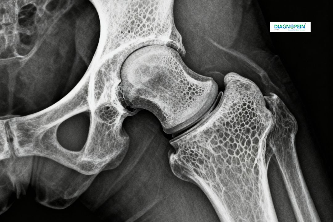
Parameters Measured in X-Ray Hip Joint One View
The X-Ray Hip Joint One View captures key anatomical structures including:
-
Femoral head and neck alignment
-
Acetabulum (hip socket) clarity
-
Joint space width and symmetry
-
Detection of fractures, calcification, or soft tissue swelling
-
Bone density and surface irregularities
These parameters help doctors evaluate bone health and detect any degenerative or traumatic changes effectively.