Why X-Ray Heel Lateral View is Important
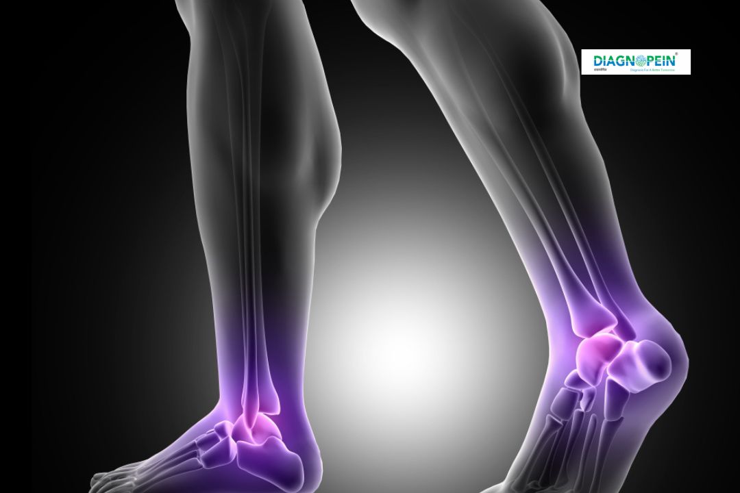
The heel supports body weight and absorbs shock during walking or running. Any damage or structural variation can cause continuous pain and discomfort. The X-Ray Heel Lateral View plays an important role in diagnosing such conditions. At Diagnopein in Karad, doctors rely on this test to obtain detailed insights into:
-
Heel fractures and bone spurs
-
Signs of calcaneal infections or tumors
-
Degenerative bone diseases
-
Structural deformities or post-trauma conditions
-
Plantar fascia or soft tissue abnormalities
Early detection of these conditions allows for better treatment planning and faster recovery. By identifying even minimal cracks or changes, X-Ray Heel Lateral View ensures precision in orthopedic and podiatric diagnoses.
Benefits of X-Ray Heel Lateral View
Getting an X-Ray Heel Lateral View at Diagnopein, Karad, offers several patient and diagnostic benefits:
-
Quick and painless procedure
-
High image clarity for bone evaluation
-
Detects hidden heel fractures and bone stress
-
Helps monitor healing progression post-surgery
-
Affordable and widely available diagnostic option
-
No need for special preparation or fasting
This examination is particularly beneficial for athletes, elderly patients, and individuals experiencing persistent heel discomfort. Since Diagnopein in Karad uses modern X-ray imaging systems, patients receive sharper images with reduced radiation exposure.
How the X-Ray Heel Lateral View is Performed
The procedure for this test is straightforward. At Diagnopein Diagnostic Center in Karad, the following steps are carried out:
-
Patients are asked to remove footwear and metallic objects.
-
The technician positions the patient either standing or lying on the X-ray table.
-
The heel is adjusted laterally to capture the desired side view.
-
The X-ray beam is directed horizontally to produce a clear lateral image of the calcaneus.
-
The image is immediately processed and reviewed by the radiologist for detailed interpretation.
The entire process typically takes 5–10 minutes, after which the digital reports and images are available for consultation. This efficient procedure ensures accurate results with minimal waiting time.
Parameters and Diagnostic Insights
During an X-Ray Heel Lateral View at Diagnopein Karad, several parameters are studied by doctors and radiologists:
-
Bone density and structure of the calcaneus
-
Fracture lines or bone defects
-
Presence of bone spurs or cysts
-
Relationship between heel bone and talus
-
Evaluation of soft tissue thickness around the heel
These insights help the physician design a customized treatment plan, whether it’s physical therapy, medication, or orthopedic support.
Why Choose Diagnopein in Karad for X-Ray Heel Lateral View
Diagnopein is known in Karad for advanced diagnostic imaging, expert radiologists, and modern technology ensuring reliable reports. The center emphasizes accuracy, patient comfort, and safety. Minimal radiation exposure and affordable rates make it the preferred choice for heel and ankle imaging.
Patients receive instant digital reports, and doctors can access high-resolution scans for better diagnostic precision. With expert staff, Diagnopein Karad stands as a trusted diagnostic solution for all X-ray and imaging needs.