Why X-Ray Both Heels AP/LAT is Done
The X-Ray Both Heels AP/LAT helps detect underlying causes of heel discomfort or trauma. It is particularly useful for identifying fractures, infections, or abnormal bone alignment. If you experience persistent heel pain, swelling, or difficulty walking, this X-ray can provide valuable diagnostic information.
Common reasons for X-Ray Both Heels AP/LAT include:
-
Heel bone or calcaneal fractures
-
Bone spurs or plantar fasciitis
-
Arthritis or degenerative changes
-
Heel deformities or alignment issues
-
Post-surgical follow-up and recovery assessment
This test aids in differentiating between conditions that may appear similar but require different treatment approaches.
Importance and Benefits of X-Ray Both Heels AP/LAT
Accurate evaluation of heel bone structure is critical for proper diagnosis and treatment. The X-Ray Both Heels AP/LAT provides detailed images of both heels from different angles, giving doctors a complete view of bone health and alignment.
Key benefits:
-
Quick, affordable, and non-invasive
-
Detects even small or hidden heel fractures
-
Helps in assessing bone density and structure
-
Guides orthopedic and rehabilitation treatment plans
-
No special preparation or fasting required
At Diagnopein karad, radiologists interpret your images accurately using digital X-ray technology, minimizing radiation exposure while improving precision.
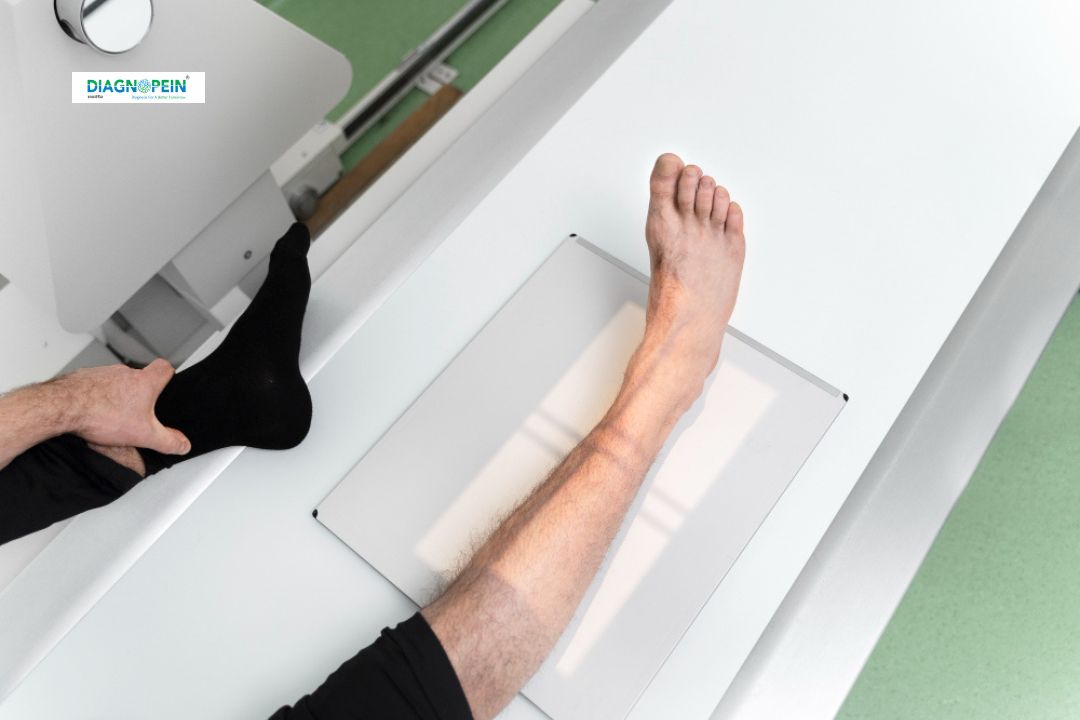
How the X-Ray Test is Performed
The X-Ray Both Heels AP/LAT procedure is simple and safe. A trained radiology technician will position you standing or sitting with your feet placed correctly on the X-ray plate.
Steps of the process:
-
You may be asked to remove footwear or metallic objects.
-
The technician captures two views: AP (front view) and LAT (side view).
-
The entire process takes less than 10 minutes.
-
The X-ray images are reviewed by a radiologist for diagnostic interpretation.
Parameters assessed:
-
Bone density and joint space
-
Calcaneal alignment and shape
-
Signs of fracture, erosion, or bone spur
-
Soft tissue changes
After your test, results are made available quickly through digital reports for your consulting doctor.
Why Choose Diagnopein Panvel for X-Ray Both Heels AP/LAT
At Diagnopein Panvel, we prioritize accuracy, safety, and comfort. Our imaging center uses modern diagnostic tools and follows strict radiation-safety protocols.
What makes us special:
-
Latest digital X-ray machines for sharp and precise imaging
-
Experienced radiologists with orthopedic expertise
-
Fast reporting and online access to results
-
Hygienic, patient-friendly environment
Diagnopein is recognized as one of the most trusted diagnostic centers in Panvel for X-Ray Both Heels AP/LAT, serving both routine and emergency patients efficiently.