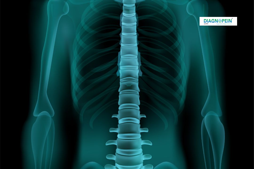
Why X-Ray Apicogram is Important
The X-Ray Apicogram plays a significant role in dental and orthopedic evaluations. It allows clinicians to inspect hard-to-see areas around tooth roots and jawbones, ensuring the detection of small cysts, lesions, or periapical infections before they become severe. In Karad, Diagnopein has enabled both dental and ENT professionals to make quick and accurate clinical decisions with X-Ray Apicogram data.
Additionally, it is crucial for diagnosis after trauma, during follow-up of root canal treatments, and in detecting chronic bone defects. This imaging tool also aids orthodontists in evaluating bone density and structure prior to surgeries or orthodontic procedures.
Benefits of X-Ray Apicogram
Choosing an X-Ray Apicogram at Diagnopein in Karad offers multiple advantages, including:
-
Precise Detection: Helps identify minute infections or bone loss not visible through standard dental X-rays.
-
Enhanced Treatment Planning: Enables dentists to perform well-informed surgeries, root canal treatments, or implants.
-
Non-invasive and Safe: Uses a very low radiation dose, ensuring patient safety.
-
Quick and Comfortable: The imaging process is painless and completed within minutes.
-
High Diagnostic Value: Useful for doctors in both dental and orthopedic setups for jawbone and skeletal evaluations.
At Diagnopein Karad, our modern radiology systems ensure that your X-Ray Apicogram produces high-resolution images with maximum clarity, supporting better clinical outcomes.
Procedure and Testing Parameters
The procedure for X-Ray Apicogram at Diagnopein in Karad is simple and efficient. The patient is positioned comfortably, and the targeted area is aligned with the X-ray beam. A small film or digital sensor is placed to capture the image. The process takes only a few minutes and does not require any fasting or preparation.
Testing parameters include:
-
Position and angle of X-ray beam alignment.
-
Exposure time (kept minimal for safety).
-
Focus area adjustment for dental roots or bone apex.
-
Contrast adjustments for optimal visualization.
Our highly trained radiology technicians ensure precise imaging while maintaining strict safety protocols. The results are instantly available for clinician review or referral to your dentist or orthopedic specialist.
X-Ray Apicogram Arthrography with Volume Imaging
In advanced cases, X-Ray Apicogram Arthrography may be performed to visualize joints and soft tissues around the bone using a minimal amount of contrast medium. This specialized approach provides volume-based imaging, enabling detailed study of joint structures, cartilage, and surrounding tissues.
Diagnopein in Karad is equipped with the latest radiographic technologies to perform Arthrography with volume imaging safely, ensuring superior diagnostic details without discomfort to the patient. It is especially beneficial for detecting joint cysts, injuries, and inflammation in the maxillofacial or orthopedic regions.
Why Choose Diagnopein in Karad
-
Experienced radiologists and certified technicians.
-
Latest-generation digital X-ray infrastructure.
-
Fast reporting and patient-friendly environment.
-
Affordable pricing for all diagnostic tests.
-
Trusted choice for X-Ray, CT, MRI, and USG in Karad.
Whether you need an X-Ray Apicogram for dental, orthopedic, or surgical reasons, Diagnopein in Karad ensures accurate imaging and timely diagnosis to support your health journey.


