Importance of X-Ray Ankle AP/LAT/Mortise
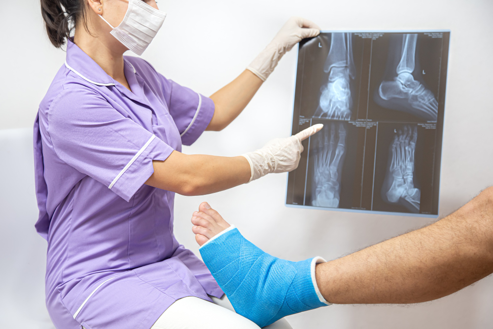
An X-Ray Ankle AP/LAT/ Mortise is a vital tool in orthopedic and radiological assessment. These three views are crucial because they allow visualization of the bony structure from multiple angles. The Mortise view, in particular, provides a detailed image of the ankle joint space, helping detect small fractures that are not visible in standard X-rays.
Some of the key reasons this test is important include:
-
Detecting bone fractures and joint dislocations
-
Evaluating ligament damage and joint alignment
-
Assessing degenerative changes due to arthritis
-
Guiding orthopedic surgical planning and post-operative follow-up
At Diagnopein, Karad, our radiology experts use state-of-the-art equipment to capture accurate details needed for precise reports. This ensures faster diagnosis, efficient treatment, and better recovery outcomes for patients.
Benefits of X-Ray Ankle Arthrography
X-Ray Ankle Arthrography is an enhanced diagnostic study where a special contrast dye is injected into the ankle joint before imaging. This helps highlight soft tissue structures such as ligaments, cartilage, and joint capsules that might not appear clearly on regular X-rays.
The main benefits include:
-
Detailed visualization of joint surfaces
-
Early detection of cartilage damage and joint diseases
-
Identification of subtle tears, inflammation, or fluid accumulation
-
Quick and painless imaging with immediate results
For patients in Karad, our Diagnopein center offers both routine and arthrographic X-Ray procedures under hygienic and comfortable conditions. Our skilled technicians ensure minimal discomfort during the procedure while maintaining high safety and precision standards.
Testing Procedure and Parameters
The procedure for X-Ray Ankle AP/LAT/Mortise at Diagnopein, Karad, is simple and efficient. The patient is asked to remove footwear and any metallic objects near the ankle area. The technician positions the foot in specific angles to capture AP, Lateral, and Mortise views.
Testing steps:
-
Patient positioning for AP view (foot flat, beam centered on the ankle joint).
-
Lateral view with side positioning to assess depth and alignment.
-
Mortise view at a 15–20-degree rotation for joint space visibility.
-
In case of Arthrography, a contrast agent is gently injected for enhanced imaging.
Parameters assessed:
-
Bone alignment and joint congruence
-
Presence of fractures or bone chips
-
Joint space width and cartilage integrity
-
Ligamentous attachment and fluid accumulation
Each X-ray is interpreted by a qualified radiologist, providing a detailed report to your referring specialist for diagnosis and treatment planning.
Why Choose Diagnopein in Karad
Diagnopein in Karad is equipped with advanced digital radiography systems ensuring superior image clarity and reduced radiation. Our trained technologists and experienced radiologists deliver prompt and accurate results. Whether it’s an emergency injury or a follow-up evaluation, our goal is to provide top-quality diagnostic imaging with compassion and professionalism.