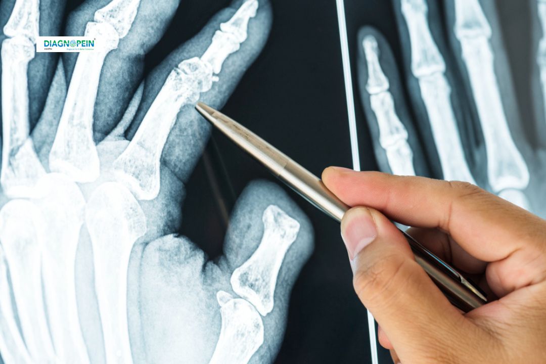
Why Wrist AP/Lat. X-ray is Important
Wrist injuries are common due to falls, sports activities, or workplace trauma. Quick and accurate diagnosis via wrist AP/Lat. X-rays is essential to plan effective treatment and prevent complications. These imaging views help doctors:
-
Detect even minor wrist fractures
-
Assess alignment after trauma
-
Reveal signs of arthritis or early degenerative changes
-
Evaluate congenital wrist abnormalities
-
Guide orthopedic management and surgical planning
The wrist AP/Lat. X-ray is non-invasive, fast, and provides clear visual evidence to confirm suspected wrist injury diagnosis. With early intervention, patients can recover functionality and avoid chronic pain. Most importantly, targeted imaging like AP and lateral views ensures that even subtle wrist pathologies are identified promptly for every patient, whether for acute trauma care or chronic wrist pain test.
Benefits of Wrist AP/Lat. X-Ray
Choosing a wrist AP/Lat. X-ray at Diagnopein offers several benefits:
-
High accuracy in detecting bone injuries or dislocations
-
Quick procedure—results are typically available within an hour
-
Safe technique with minimal radiation exposure
-
Guides physicians for further orthopedic treatment or surgery
-
Facilitates thorough evaluation for unexplained wrist pain, swelling, or deformity
-
Suitable for patients of all ages, including children with growth plate (epiphyseal) injuries
By utilizing both AP (anteroposterior) and lateral views, clinicians get a comprehensive 3D perspective of the wrist anatomy. These tests are invaluable for precise trauma diagnosis, chronic conditions, and for monitoring healing after treatment.
How Wrist AP/Lat. X-Ray is Performed
At Diagnopein, our protocol for wrist AP X-ray and wrist Lat. X-ray ensures maximum patient comfort and image clarity:
-
The patient’s affected wrist is positioned on the X-ray table
-
For the AP view, the palm faces up on the table for a front-to-back exposure
-
For the Lat. view, the side of the wrist is placed against the detector for a side-to-side image
-
The radiographer may use small positioning aids to keep the wrist steady
-
Patients should remove bracelets, watches, or metal accessories to avoid interference
-
The scan is quick, painless, and usually completed in 10-15 minutes
Common Parameters Evaluated
-
Bone alignment and joint spaces
-
Detection of fractures, breaks, or chips
-
Assessment of soft tissue swelling
-
Early signs of arthritis or bone disease
-
Any abnormal growth or deformity
Our center is committed to delivering superior wrist imaging and personalized care. For reliable trauma X-ray, orthopaedic wrist X-ray or routine wrist pain test, trust Diagnopein for your diagnostic needs in Karad.