Why Uterus Cervix Ovary FT Test is Important
The Uterus Cervix Ovary FT ultrasound is an essential part of women’s health assessment. It provides a clear view of the reproductive organs, helping doctors detect problems early and plan suitable treatments. This test is important for:
-
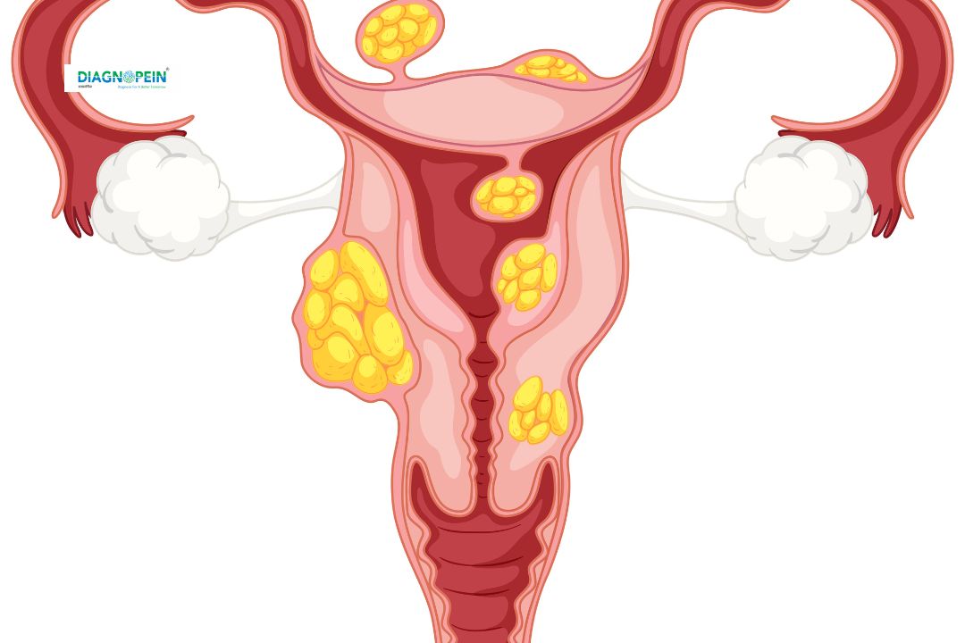
Evaluating the uterus for fibroids, polyps, or congenital abnormalities.
-
Assessing the cervix for any infection, abnormalities, or stenosis.
-
Examining the ovaries for cysts, PCOD/PCOS, or tumors.
-
Checking the fallopian tubes (FT) for blockage, fluid collection, or infections.
Regular ultrasound screening of the uterus, cervix, ovaries, and FT helps detect potential issues affecting fertility or menstrual health. Both married and unmarried women experiencing pelvic pain, abnormal bleeding, or infertility concerns may benefit from this test.
Benefits of Uterus Cervix Ovary FT Test
There are multiple clinical and health benefits of performing the Uterus Cervix Ovary FT ultrasound. Some of the major benefits include:
-
Non-invasive procedure: It is a safe, painless, and radiation-free imaging technique.
-
Early detection: Helps identify ovarian cysts, fibroids, endometriosis, and tubal blockages in the early stages.
-
Fertility evaluation: Supports fertility assessment by monitoring ovulation and follicle development.
-
Treatment guidance: Helps in planning medical or surgical treatment for uterine or ovarian issues.
-
Menstrual health monitoring: Useful in evaluating abnormal or heavy menstrual bleeding causes.
Overall, this scan supports a comprehensive view of female reproductive wellbeing and plays a vital role in maintaining gynecologic and fertility health.
How the Uterus Cervix Ovary FT Test is Done
The Uterus Cervix Ovary FT ultrasound is a quick and comfortable process typically done in two methods—Transabdominal and Transvaginal (TVS) ultrasound, depending on the patient’s clinical requirement.
Procedure Steps:
-
The patient is asked to drink water before the test to fill the bladder (for transabdominal scan).
-
During the scan, a gel is applied on the abdomen or a transducer is gently inserted (for TVS) to capture clear images.
-
The radiologist examines the uterus, cervix, ovaries, and fallopian tubes (FT) for any structural or functional changes.
-
The entire process usually takes 15–20 minutes and results are reviewed by a gynecologist or fertility specialist.
Parameters Evaluated
-
Uterine size, position, and endometrial thickness
-
Cervical length and texture
-
Ovarian size, cyst presence, and follicular count
-
Fallopian tube patency and fluid collection