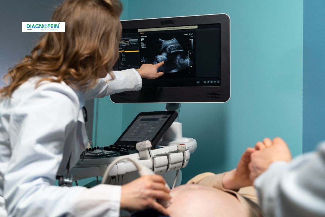
Why Choose USG Guided Intervention – FNAC?
Choosing USG guided FNAC offers numerous clinical advantages over traditional methods. The ultrasound-guided approach ensures high accuracy in needle placement and minimizes the risk of sample contamination or bleeding.
Key reasons to prefer USG-guided intervention:
-
Accurate Sampling: Real-time visualization ensures that the needle precisely targets the suspicious area.
-
Minimally Invasive: No incisions or stitches are required, making it safer and less painful.
-
Quick Results: Samples collected through ultrasound-guided FNAC are sent for cytology, with faster turnaround times for reports.
-
Safe for All Age Groups: The procedure has minimal side effects or post-procedure complications.
-
Affordable: As a cost-effective diagnostic choice, USG guided FNAC test offers advanced results at a reasonable price.
This technique is especially helpful for identifying malignancies, differentiating cystic from solid lesions, and monitoring previously treated conditions.
How the USG Guided FNAC Test is Done
The USG guided FNAC procedure is usually performed by an experienced radiologist or cytopathologist using high-resolution ultrasound equipment. Below is the step-by-step process:
-
Preparation: The patient is positioned comfortably depending on the biopsy site (such as neck, breast, or abdomen). No fasting or anesthesia is usually required.
-
Localization: The radiologist scans the target region using ultrasound to visualize the exact location of the lesion.
-
Needle Insertion: Under continuous ultrasound guidance, a thin and sterile needle is inserted into the lesion to draw a small amount of fluid or tissue sample.
-
Sample Collection: The specimen is placed on glass slides for microscopic examination.
-
Post-Procedure Care: The site is cleaned and bandaged. Patients can resume normal activities immediately after the procedure.
The USG guided FNAC test usually takes 15–30 minutes and causes minimal discomfort. Patients may only experience mild tenderness at the puncture site, which resolves within a day.
Parameters and Reporting
Cytological parameters examined in ultrasound-guided FNAC include:
-
Cell type analysis to identify malignancy or benign nature.
-
Cytoplasmic and nuclear features for detailed morphology.
-
Presence of necrosis, inflammation, or atypia for accurate categorization.
-
Correlation with ultrasound findings for better clinical interpretation.
Results from USG guided FNAC are typically available within 24–48 hours. The report helps the physician decide on further investigations or treatment options such as surgery, medication, or follow-up imaging.
Benefits of USG Guided FNAC
-
High diagnostic accuracy even for small or deep-seated lesions.
-
Reduced procedure risk with minimal pain and complications.
-
Helps in early cancer diagnosis and timely treatment.
-
Can be repeated if needed without additional risk.
-
Provides real-time imaging control for needle placement.
Whether it’s for thyroid FNAC, breast FNAC, or lymph node cytology, the procedure provides critical insights that guide patient care effectively.


