Importance of USG Small Parts
The USG Small Parts examination is an essential component in modern diagnostic medicine. It allows detailed evaluation of small organs that are difficult to assess through other imaging modalities like CT or MRI due to their size or location.
Key reasons why USG Small Parts is important include:
-
Detects thyroid nodules, lymph node enlargement, or salivary gland disorders.
-
Evaluates breast lesions and helps differentiate between benign and malignant findings.
-
Assists in scrotal imaging for detecting varicocele, testicular masses, or inflammation.
-
Identifies soft tissue swellings, lipomas, or cysts under the skin surface.
With this technology, physicians can diagnose diseases early, track treatment response, and decide whether further imaging or biopsy is needed.
Benefits of USG Small Parts
The USG Small Parts scan offers multiple patient-centered benefits:
-
Non-invasive and radiation-free – Safe for repeated use, including pediatric and elderly patients.
-
Real-time imaging – Provides dynamic observation of tissues and blood flow.
-
Immediate results – Enables quick diagnosis and medical decision-making.
-
Cost-effective – More affordable than MRI or CT scans for soft tissue evaluations.
-
Guidance for fine-needle aspiration or biopsy – Improves sampling accuracy.
At Diagnopein, our high-resolution ultrasound systems ensure superior image clarity and precision. All scans are conducted by experienced radiologists following standardized protocols for each region examined.
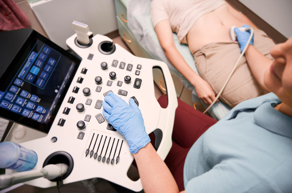
How USG Small Parts Testing Is Done
The procedure for USG Small Parts is simple and painless. No major preparation is required, making it convenient for both patients and doctors.
Testing Procedure Steps:
-
The patient is comfortably positioned depending on the area being examined.
-
A layer of ultrasound gel is applied to allow smooth transmission of sound waves.
-
The radiologist moves a handheld transducer over the target area.
-
Images are captured and analyzed in real time on the ultrasound monitor.
The test usually takes 15–30 minutes. After the scan, the images are interpreted by a radiologist who provides a detailed report highlighting normal and abnormal findings.
Parameters Evaluated in USG Small Parts
During the USG Small Parts study, several vital parameters are assessed:
-
Organ size, shape, and contour – for structural abnormalities
-
Internal echo texture – for identifying cystic, solid, or mixed lesions
-
Presence of calcifications or nodules
-
Vascularity patterns using color Doppler
-
Lymph node characteristics – size, borders, and hilum visibility
These parameters help the doctor determine if the tissue is healthy or affected by disease. At Diagnopein, detailed imaging protocols provide comprehensive evaluations ensuring diagnostic confidence.
Why Choose Diagnopein for USG Small Parts
Diagnopein combines cutting-edge ultrasound equipment, experienced radiologists, and patient-friendly service. Our focus on accuracy, comfort, and clarity ensures every USG Small Parts scan delivers confidence and reliability. With same-day reporting and expert consultations, patients can quickly take the next step toward treatment.
We make sure that every USG Small Parts examination follows quality imaging standards for efficient, accurate, and safe results.