What is USG Local Part?
USG Local Part (Ultrasound Localized Region) focuses on a specific part of the body such as the neck, upper limb, thigh, scrotum, or any localized swelling. The scan helps doctors visualize structures beneath the skin with clarity and precision. It is often prescribed when a patient experiences localized discomfort, suspected cysts, or swelling in a particular body region.
Unlike other imaging methods, Ultrasound Local Part uses sound waves instead of radiation, making it ideal for children and pregnant women. The exam is widely used across hospitals and diagnostic centers because it is quick, safe, and provides immediate results.
Common Areas Scanned Using USG Local Part:
-
Neck or cervical region
-
Upper or lower limbs
-
Shoulder or joint areas
-
Scrotum or groin
-
Localized soft tissue swellings
Why USG Local Part is Done
Doctors recommend a USG Local Part examination to investigate specific complaints such as localized pain, inflammation, or lumps. The test helps identify early signs of infections, fluid collection, abscesses, cysts, or soft tissue injuries. In many clinical cases, this targeted ultrasound provides instant insight into the problem area without the need for more complex imaging.
This imaging study is also used to monitor recovery from trauma or postoperative conditions, evaluate blood flow in small vessels, and assist in guided procedures like aspiration or biopsy. The Ultrasound Local Part test offers high-resolution images that help clinicians make accurate diagnoses and treatment plans.
Key Reasons for USG Local Part Scan:
-
To detect abscesses or cysts
-
To assess localized pain or swelling
-
To evaluate soft tissue injuries
-
To guide minor procedures or injections
-
To monitor healing or follow-up treatment
Benefits of USG Local Part
The USG Local Part test offers multiple clinical and patient-focused advantages that make it an essential diagnostic tool in modern healthcare.
Major Benefits:
-
Non-invasive and painless: No needles or incisions are involved.
-
No radiation exposure: Safe for pregnant women and all age groups.
-
Real-time imaging: Provides dynamic visualization of moving tissues.
-
Cost-effective and accessible: Widely available and affordable compared to MRI or CT.
-
Instant reporting: Results are often available immediately after scanning.
For patients with localized health issues, Ultrasound Local Part offers fast and accurate insight, allowing early detection and better treatment outcomes.
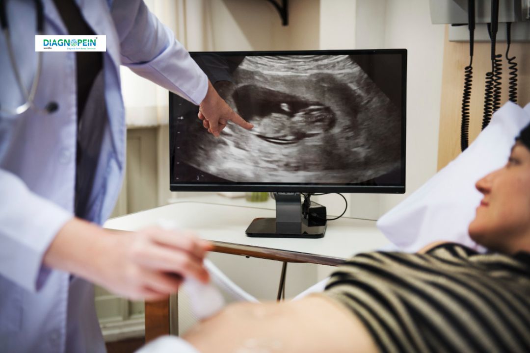
How the USG Local Part Test is Performed
The procedure for USG Local Part is simple, quick, and non-invasive. No special preparation is required for most body areas.
-
The patient is positioned comfortably depending on the body part to be examined.
-
A gel is applied on the skin to help sound waves pass smoothly.
-
The radiologist moves a handheld probe (called a transducer) over the target area.
-
The images are displayed in real time on the ultrasound monitor.
-
After the scan, the gel is wiped off, and results are reviewed by the doctor.
The entire scan usually takes 10–20 minutes. Patients can resume their normal activities immediately afterward.
Scanning Parameters:
-
Frequency range: 5–12 MHz
-
Mode: B-mode 2D grayscale imaging
-
Depth adjustment based on the scanned area
-
Real-time Doppler (if vascular flow is evaluated)
Summary
USG Local Part is a safe, quick, and affordable imaging test suited for evaluating localized areas of concern such as pain, swelling, or soft tissue injuries. It helps doctors in precise diagnosis through real-time imaging without radiation. The test is widely available in diagnostic centers and hospitals, promising accuracy and comfort for patients.
For the best diagnostic outcomes, choose an advanced imaging facility offering high-quality Ultrasound Local Part services performed by qualified radiologists using modern ultrasound machines.