Why USG Fetal Echo is Important
Performing a USG Fetal Echo Test helps in identifying congenital heart diseases such as septal defects, valve abnormalities, and rhythm disorders at an early stage. Pregnant women are often advised to undergo a Fetal Echocardiography Scan if they have pre-existing conditions like diabetes, a history of congenital heart disease, or abnormal findings on a routine ultrasound.
Key indications for USG Fetal Echo in pregnancy include:
-
Family history of congenital heart defects
-
Maternal infections such as rubella
-
Exposure to medications or substances that can affect fetal development
-
Abnormal fetal heart rate or rhythm
-
Chromosomal abnormalities detected through screening
This test offers reassurance to expecting parents and prepares the healthcare team for proper management if any issues are found.
Benefits of USG Fetal Echocardiography
Some of the major benefits of USG Fetal Echo Scan include:
-
Early and accurate detection of fetal heart abnormalities
-
Non-invasive and completely safe for both mother and baby
-
Provides essential information for planning delivery and postnatal treatment
-
Helps specialists recommend intervention or revision of care plan as needed
-
Offers peace of mind to parents with detailed heart assessment of the fetus
By identifying conditions before birth, Fetal Echo Ultrasound supports better outcomes for both baby and mother. It is one of the most reliable diagnostic tools for evaluating fetal cardiac health.
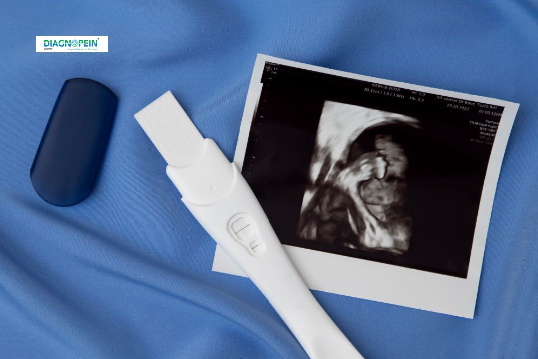
How the USG Fetal Echo Test is Performed
The Fetal Echocardiography Procedure is similar to a standard ultrasound. A trained radiologist or fetal medicine specialist performs the scan using a high-frequency transducer.
During the test:
-
The mother lies comfortably on the examination table.
-
A gel is applied over the abdomen to ensure good contact between the transducer and the skin.
-
The ultrasound probe is moved gently to capture different heart views.
-
Images and blood flow are examined using Doppler technology.
The entire process of USG Fetal Echo usually takes 30–45 minutes. It is painless, requires no preparation, and produces immediate results for preliminary evaluation.
When to Get a USG Fetal Echo Done
Doctors may recommend USG Fetal Echo in pregnancy in the following situations:
-
History of congenital heart disease in family
-
Abnormal heart rate noticed on routine fetal scan
-
Mother having diabetes, lupus, or thyroid disorders
-
In-vitro fertilization (IVF) pregnancy
-
Increased nuchal translucency (NT) measurement
Regular prenatal screening with USG Fetal Echocardiography not only ensures fetal well-being but also helps healthcare providers in planning a safe childbirth.