Why USG Doppler of Upper/Lower Limb Venous is Needed
A USG Doppler of Upper or Lower Limb Venous system is essential for evaluating vascular health and venous circulation. It helps identify abnormalities such as deep vein thrombosis, varicose veins, vascular obstruction, venous insufficiency, and other blood flow irregularities. For patients with leg swelling, pain, or post-surgery vein concerns, this test is a key diagnostic tool.
Doctors use the results of a venous Doppler ultrasound to decide on further treatments like anticoagulant medication, vein surgery, or compression therapy. The test provides accurate information about blood flow direction and valve efficiency inside the veins, helping ensure timely medical intervention and preventing complications like pulmonary embolism.
Benefits of USG Doppler Venous Examination
The USG Doppler Upper/Lower Limb Venous scan offers multiple advantages in clinical practice. It provides real-time imaging of venous blood flow without exposing the patient to radiation. The test is painless, quick (usually 20–30 minutes), and delivers instant results for better vascular diagnosis.
Key benefits include:
-
Early detection of deep vein thrombosis (DVT) or vein blockages.
-
Monitoring post-surgical venous function in limbs.
-
Identifying varicose veins and venous reflux.
-
Guiding vascular surgeons in treatment planning.
-
Preventing serious conditions such as pulmonary embolism or chronic leg ulcers.
Because the USG Doppler Venous study is safe during pregnancy and suitable for repeated use, it is preferred over invasive tests like venography. The results are usually interpreted by a radiologist specializing in vascular imaging.
How the USG Doppler Venous Test is Done
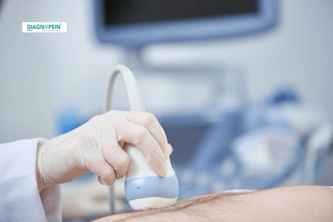
During a USG Doppler Upper/Lower Limb Venous R/L study, the patient lies comfortably on an examination table. A water-based gel is applied over the limb area to allow smooth transducer movement. The radiologist systematically moves the Doppler probe along the veins of the upper or lower limb while monitoring real-time blood flow patterns on a screen.
The transducer sends sound waves into the body, and their reflections create detailed images of vein structure and blood velocity. The process is non-invasive and takes 20–40 minutes depending on the area being scanned (right/left, upper/lower limb).
After the scan, the radiologist generates a color Doppler report showing vein compressibility, flow pattern, and any presence of thrombus or obstruction. No specific fasting or preparation is needed before the test.
Important Parameters Evaluated:
-
Venous flow pattern and velocity
-
Vein wall thickness
-
Presence of thrombus or varicose changes
-
Valve efficiency and reflux signs
-
Comparison between right and left limb flow dynamics
5. Summary and Diagnostic Value
A USG Doppler Upper/Lower Limb Venous test plays a crucial role in diagnosing vascular and venous disorders early. It helps clinicians manage chronic venous disease, swelling, DVT, and circulation disorders accurately without invasive procedures. This test is strongly recommended for patients with leg pain, swelling, or suspected vein blockage for timely diagnosis and treatment.