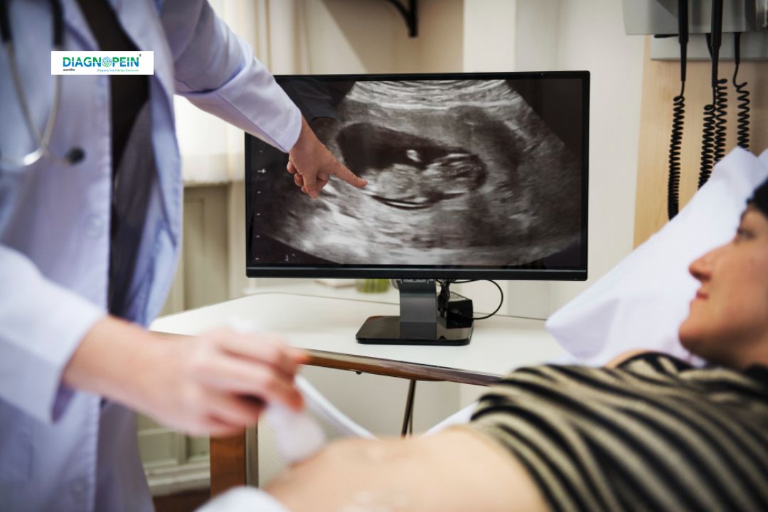
Why USG Chest?
USG Chest is preferred for its quick, radiation-free, and precise diagnostic capabilities. It allows healthcare providers to assess chest abnormalities dynamically and guide clinical decision-making in real-time.
This imaging technique helps identify:
-
Pleural effusion (fluid around the lungs)
-
Lung consolidation (infection or pneumonia)
-
Diaphragmatic paralysis or movement disorders
-
Pneumothorax (collapsed lung)
-
Chest wall or mediastinal masses
When early diagnosis is crucial, USG Chest provides instant insights into lung and pleural conditions, helping in rapid management of respiratory diseases. It is particularly beneficial in ICU settings and for patients who cannot be easily shifted to radiology for X-ray or CT scans.
Benefits of USG Chest
There are several advantages of performing a USG Chest over conventional imaging, making it a valuable diagnostic tool in chest medicine.
-
No Radiation Exposure: Uses sound waves, making it completely safe.
-
Real-Time Visualization: Allows doctors to see live movement of the lungs, pleura, and diaphragm.
-
Guided Procedures: Assists in procedures like pleural tapping, biopsies, or drainage with precision.
-
Portable & Bedside Use: Ideal for ICU or critical care patients.
-
Cost-Effective: More affordable compared to CT or MRI imaging.
Clinicians prefer USG Chest because of its ability to detect even minimal liquid collections and guide therapeutic interventions. It is a valuable adjunct for continuous monitoring in respiratory patients.
How USG Chest Test is Done
The USG Chest procedure is simple, painless, and usually takes 10–20 minutes.
-
Patient Positioning: The patient may be asked to sit upright or lie on their back or side.
-
Application of Gel: A water-based gel is applied to the chest area to ensure clear sound wave transmission.
-
Ultrasound Probe: The radiologist moves a transducer (probe) over the chest and back to capture detailed images.
-
Image Interpretation: The live images are analyzed to identify abnormalities like fluid, inflammation, or collapse.
No special preparation or fasting is required for this test. Results are typically available immediately, enabling faster diagnosis and timely treatment decisions.
Parameters and Findings in USG Chest
During a USG Chest, several diagnostic parameters are assessed for clarity and accuracy of chest health.
Key parameters include:
-
Pleural fluid depth and characteristics
-
Diaphragmatic motion
-
Lung sliding and artifacts (A-lines, B-lines)
-
Parenchymal patterns (consolidation, atelectasis)
-
Presence of lesions, cysts, or fibrosis
These parameters help radiologists detect both major and subtle chest abnormalities that might not be visible on a chest X-ray. USG Chest findings play a crucial role in critical care management, guiding thoracentesis, and evaluating disease progression.
Conclusion
USG Chest is a safe, fast, and highly informative diagnostic investigation for evaluating chest and lung conditions. With zero radiation and bedside usability, it remains one of the most efficient methods for assessing chest diseases in both hospital and outpatient settings.
If you’re experiencing chest pain, breathing difficulty, or suspected lung infection, consult your physician for a USG Chest Scan today for early detection and better care outcomes.


