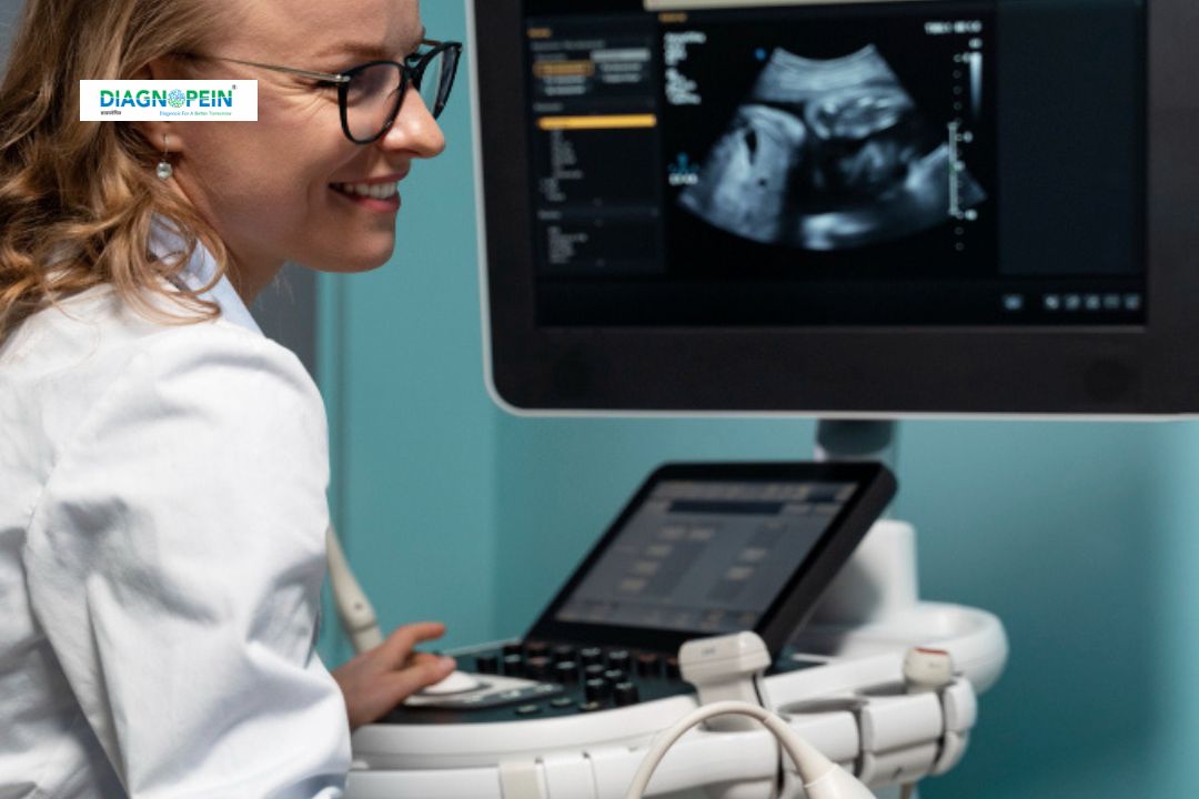
Why Choose USG 3D Image?
The decision to undergo USG 3D Image scanning offers multiple benefits over conventional imaging methods. The clarity and precision of 3D imaging allow for enhanced visualization of organs and soft tissues, contributing to better medical evaluation and treatment planning.
Key reasons to choose USG 3D Image test:
-
Provides realistic 3D visuals of fetus, liver, kidney, or uterus.
-
Helps in early detection of structural or growth abnormalities.
-
Completely safe and non-invasive imaging technique.
-
Offers better insights into organ function and tissue details.
-
Ideal for prenatal checkups, gynecological studies, and abdominal scans.
Choosing USG 3D ultrasound also helps healthcare providers identify subtle changes that might go unnoticed in standard 2D images. The advanced software used in our lab enhances depth resolution and volume measurement accuracy.
Benefits of USG 3D Image Test
USG 3D Image has revolutionized the field of diagnostic scanning through its enhanced visualization and improved interpretive detail. Both patients and doctors benefit from a clearer, more accurate representation of internal anatomy.
Major advantages of USG 3D Image include:
-
High-resolution 3D visuals: Offers more lifelike imaging for better understanding.
-
Improved diagnostic accuracy: Detects minute structural changes in organs.
-
Fetal monitoring: Provides detailed visualization of fetal movements, position, and growth.
-
Pain-free examination: Completely non-invasive and does not involve radiation exposure.
-
Quick and convenient: Delivers instant results for immediate analysis.
Clinicians rely on 3D USG scanning to monitor high-risk pregnancies, assess congenital anomalies, and evaluate reproductive health. The entire process is designed to prioritize patient comfort, accuracy, and diagnostic excellence.
How Is USG 3D Image Test Done?
The USG 3D Image test is simple, safe, and comfortable. It typically takes around 20–30 minutes, depending on the area being scanned.
Step-by-step procedure:
-
The patient is positioned comfortably on an examination bed.
-
A layer of clear gel is applied to the scanning area to ensure smooth soundwave transmission.
-
The radiologist uses a transducer to capture multiple 2D image slices.
-
These slices are processed by the ultrasound machine to create a complete 3D image volume.
-
The results are displayed on a high-definition monitor for detailed evaluation.
The procedure requires no fasting or special preparation unless advised by your doctor. For expecting mothers, 3D ultrasound scanning is an emotional and informative experience, providing realistic images of the baby inside the womb.
Parameters and Image Quality
During USG 3D scanning, several technical parameters determine image clarity and accuracy:
-
Frequency range: Usually between 2–10 MHz based on organ depth.
-
Volume data acquisition: Combines multiple 2D images into one 3D dataset.
-
Contrast and brightness control: Ensures detailed differentiation between tissues.
-
Depth resolution: Provides multi-plane and volumetric analysis.
Using advanced ultrasound machines, our diagnostic team ensures each USG 3D Image meets the highest standards of clinical precision and imaging sharpness.


