Why Sonography Lower Abdomen is Recommended
Doctors usually recommend Sonography Lower Abdomen when there are symptoms such as persistent abdominal pain, frequent urination, irregular menstrual cycles, or swelling in the lower stomach region. The scan helps detect the cause of these symptoms and provides diagnostic clarity.
In men, this test often focuses on the urinary bladder and prostate gland, helping in early detection of enlargement or infections. In women, it helps evaluate the uterus, fallopian tubes, and ovaries for cysts, fibroids, or any other gynecological conditions.
Lower Abdomen Ultrasound can also be suggested to monitor the position and health of internal organs after a medical treatment or surgery. It is considered one of the safest and most effective diagnostic tools available in modern imaging.
Conditions Detected:
-
Urinary bladder infections or stones
-
Prostate enlargement or inflammation
-
Uterine fibroids or cysts
-
Ovarian cysts or tumorous growths
-
Post-surgical monitoring
Benefits of Sonography Lower Abdomen
Undergoing Sonography Lower Abdomen has multiple benefits for both diagnostic and preventive care purposes.
Non-invasive and safe: Unlike CT scans or X-rays, there is no radiation exposure, making it safe for all age groups.
Quick and painless: The test is completed in 15–20 minutes with instant report generation.
Accurate detection: It provides clear imaging that supports precise diagnosis.
Cost-effective: It is affordable compared to other imaging technologies while maintaining a high diagnostic value.
Useful for both genders: Effective in diagnosing both male and female abdominal or pelvic issues.
For pregnant women, a Lower Abdomen Ultrasound may also be performed to monitor fetal development or rule out any lower abdominal complications safely.
How the Test is Performed
The Sonography Lower Abdomen test is simple and does not require complex preparation.
-
You may be advised to come with a full urinary bladder for better image clarity.
-
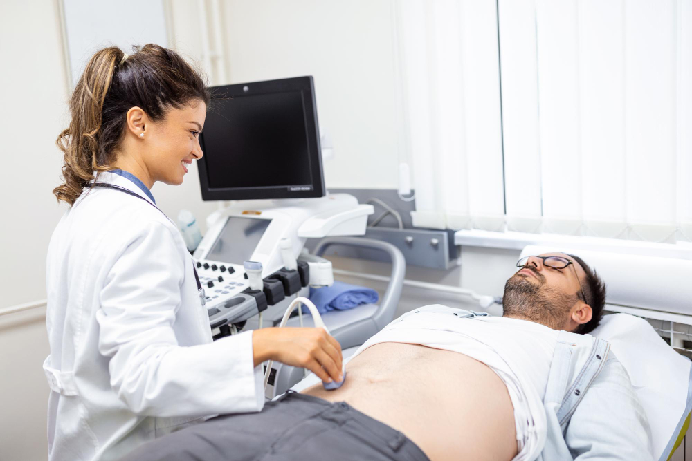
The patient lies flat on the examination table, exposing the lower abdomen.
-
A special gel is applied to the skin, which helps sound waves travel through.
-
The radiologist moves a small transducer device over the abdomen to capture images.
-
The images are displayed on a monitor and analyzed immediately.
The entire process takes around 15 to 20 minutes, and the results are interpreted by a qualified radiologist. Reports are usually available the same day, enabling the doctor to plan further care quickly.
Sonography Parameters and Report Results
When performing Sonography Lower Abdomen, several internal parameters are recorded to ensure diagnostic accuracy:
-
Size and shape of the bladder, uterus, or prostate
-
Wall thickness of abdominal organs
-
Detection of cysts, masses, fluid accumulation, or stones
-
Blood flow observations with color Doppler (when needed)
-
Presence of infection or inflammation
The final report gives detailed measurements and findings that assist doctors in diagnosis. If abnormalities are found, further imaging like CT, MRI, or specialized scans may be recommended for confirmation.
Why Choose Our Sonography Center in Karad
Our facility offers advanced Sonography Lower Abdomen imaging services using high-resolution ultrasound machines and skilled radiologists. We ensure comfortable procedures, accurate reports, and fast delivery times so patients can proceed with their treatments without delay.
Our center in Karad follows hygienic and patient-friendly protocols, ensuring each procedure delivers maximum clarity and comfort.


