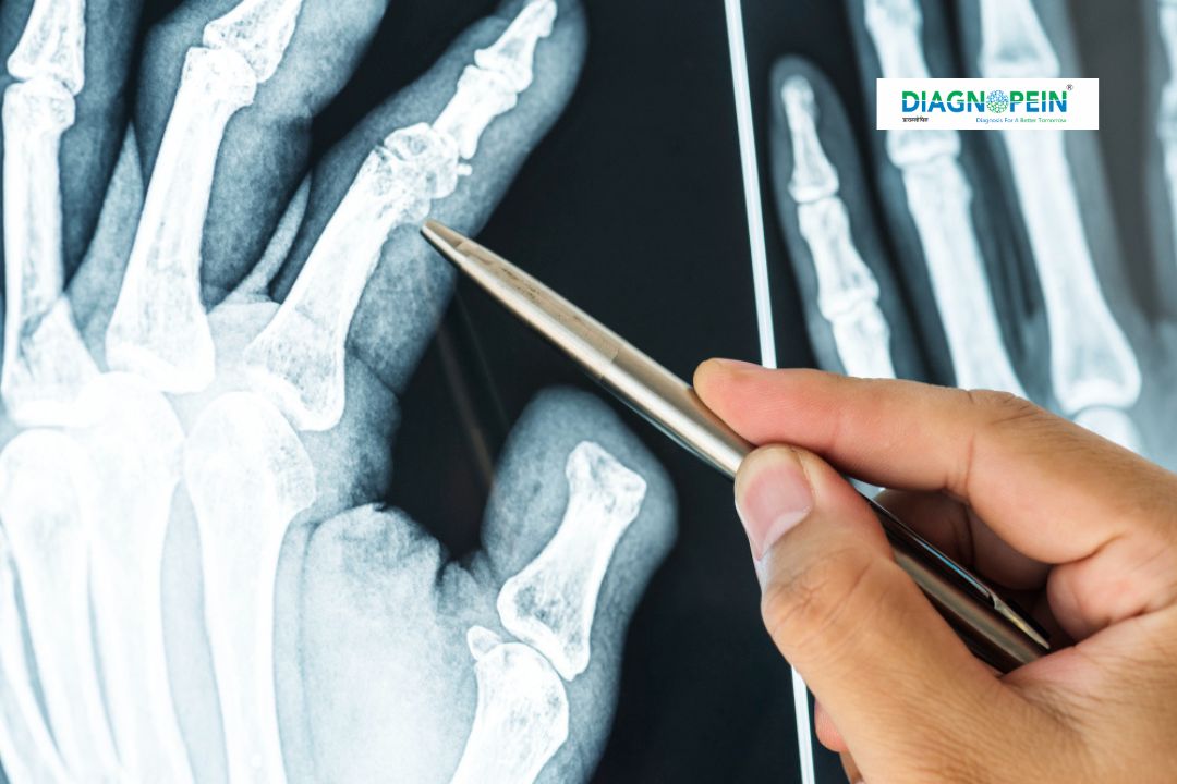
Importance of Patella AP/Lateral/Axial View
This radiographic test is crucial for evaluating several orthopedic and sports-related conditions that affect the kneecap. It helps detect and monitor:
-
Patellar fractures and displacement
-
Osteoarthritis or degenerative joint disease
-
Patellar alignment and tracking disorders
-
Post-surgical complications after knee surgery or replacement
-
Soft tissue calcifications affecting joint movement
The combination of AP (Anteroposterior), Lateral, and Axial or “sunrise” views provides a complete examination from multiple angles. These perspectives allow better visualization of the patella’s articulation with the femur, helping specialists plan correct treatment protocols or surgical interventions.
Modern imaging technologies such as OrthoScan Patella Karad and FlexiView Patella Karad enable medical professionals to detect subtle abnormalities that conventional imaging systems may overlook.
Benefits of Patella AP/LAT/Axial X-Ray
Performing a Patella AP/Lateral/Axial View X-ray offers many advantages for both patients and clinicians:
-
Accurate Diagnosis: The multi-view approach reveals both joint alignment and bone surface conditions.
-
Quick and Non-invasive: The procedure is painless, fast, and does not require anesthesia.
-
Low Radiation Exposure: Modern digital X-ray units, like those used in KneeSure X-Ray Karad, minimize radiation exposure while delivering clear, diagnostic-quality images.
-
Enhanced Treatment Planning: The results guide orthopedic experts in designing effective treatment plans, physiotherapy regimens, or surgical corrections.
-
Dedicated Post-surgical Evaluation: The post-operative condition of implants or knee reconstructions can be assessed easily through the axial and lateral views.
By integrating cutting-edge imaging platforms such as JointVision Karad, Diagnopein ensures optimal imaging outcomes while keeping patient safety as the top priority.
Procedure: How the Test is Performed
The Patella AP/LAT/Axial View is conducted by trained radiology technicians under sterile and patient-friendly conditions. The process involves minimal preparation and is completed within a few minutes.
Steps involved:
-
The patient is positioned appropriately on the X-ray table. For the AP view, the knee is extended straight while the beam passes from front to back.
-
For the Lateral view, the patient lies on the side, and the X-ray beam passes across the knee joint from side to side.
-
The Axial or Skyline view requires the knee to be flexed at a specific angle, allowing imaging of the patellofemoral joint directly from above.
-
The radiologist reviews all three images to ensure quality and clarity before interpretation.
The test requires no fasting or injections. Pregnant patients should inform the technician before imaging, as radiation exposure must be minimized during pregnancy. Once completed, digital images are stored securely in Diagnopein’s radiology system for immediate review and reporting.
Parameters and Technical Details
Key parameters considered during Patella AP/Lateral/Axial imaging include:
-
Exposure settings: Adjusted for optimal bone contrast (around 60–70 kVp).
-
Beam alignment: Centered precisely at the patella to ensure joint symmetry.
-
Patient positioning: Accurate angle alignment to avoid image distortion.
-
Radiation dose: Kept as low as possible under ALARA (As Low As Reasonably Achievable) standards.
Our expert radiology team at Diagnopein, equipped with modern imaging units such as Karad OrthoVue Patella, ensures every scan is optimized for clarity, safety, and diagnostic precision.
Why Choose Diagnopein Karad for Patella Imaging
Diagnopein combines advanced imaging infrastructure, expert technicians, and orthopedic collaboration to offer superior diagnostic accuracy. Whether it’s a detailed bone study through Patellaview 3D Karad or a quick KneeSure X-Ray Karad, patients in Karad trust our expertise in knee and orthopedic imaging. Our commitment to quality and patient care makes us a preferred destination for accurate knee evaluation and joint health imaging.


