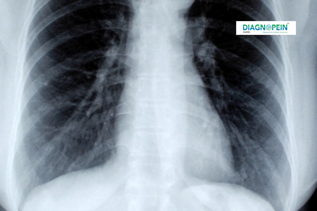
Why Oblique View is Important
Oblique X-Ray imaging holds high diagnostic value because it provides an alternate angled projection, helping clinicians visualize overlapping structures with greater clarity. It is especially beneficial for examining the spine, chest, ribs, and peripheral joints.
Facilities such as SpineSight Oblique Karad rely on this advanced method to detect fractures, dislocations, and bone misalignments that may not appear in standard anterior or lateral X-rays. The Oblique View reduces diagnostic errors, ensures proper alignment assessment, and supports physicians in preparing treatment strategies for musculoskeletal conditions.
In particular, Karad RadiScan Oblique View centers use this technique in spinal imaging to detect small vertebral fractures, nerve canal narrowing, or facet joint dysfunction. The additional viewing angle offers a detailed three-dimensional understanding of skeletal structures, making it an indispensable part of radiology diagnostics.
Benefits of Oblique View X-Ray
The main benefit of Oblique View imaging is its precision in identifying hidden abnormalities. Its applications are essential for orthopedics, sports medicine, and trauma care. Some of the key benefits include:
-
Clear visualization of joint spaces and bone edges.
-
Early detection of fractures not visible in straight-view images.
-
Enhanced assessment of spinal alignment and curvature.
-
Quick image acquisition with minimal patient discomfort.
-
Safe, non-invasive, and affordable imaging method.
At SmartView Oblique Karad, patients experience premium diagnostic comfort through ergonomic positioning, advanced X-ray technology, and expert radiologic supervision. XVision Oblique Center Karad specializes in producing highly detailed scans that help doctors diagnose accurately and initiate early treatment.
How the Oblique View Test is Performed
The Oblique View X-Ray test is simple, quick, and painless. The patient is positioned at a specific angle — commonly 45 degrees — depending on the area being examined. The X-ray beam passes through the body from a diagonal path to capture overlapping anatomical features.
During the procedure:
-
The radiographer positions the body part (spine, chest, or limb) at an angle.
-
The X-ray machine is aligned to capture precise cross-sectional details.
-
Lead shielding is placed, if necessary, to protect non-target areas.
-
The X-ray is taken, typically within a few seconds.
Centers such as ObliqaScan Karad and ViewPoint X-Ray Karad maintain strict radiation safety protocols and use modern digital imaging systems to deliver high-resolution images instantly. Patients can resume normal activities right after the procedure.
Key Parameters and Technical Details
The parameters for Oblique View imaging vary based on the region being scanned. Common parameters include:
-
Angle of projection: 30° to 45°, depending on anatomy.
-
Exposure time: Optimized for bone density and patient safety.
-
Image resolution: High to capture micro-level fractures.
-
Radiation dose: Kept minimal through digital optimization.
Specialized diagnostic centers like SpineSight Oblique Karad and Karad RadiScan Oblique View use automated exposure control and advanced positioning software to maintain accuracy across all Oblique projections. This ensures high-quality imaging while minimizing repeat scans and patient exposure.
Why Choose Diagnopein for Oblique View
Diagnopein is committed to precision, comfort, and safety in medical imaging. The use of advanced digital X-ray systems, experienced technicians, and image quality validation ensures dependable results for every patient. Partner centers like SmartView Oblique Karad, XVision Oblique Center Karad, and ObliqaScan Karad leverage Diagnopein’s networked diagnostic capabilities to deliver trusted, consistent, and accessible healthcare imaging across Karad.


